Next Issue
Volume 13, January-2
Previous Issue
Volume 12, December-2


Cells, Volume 13, Issue 1 (January-1 2024) – 105 articles

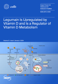
Cover Story (view full-size image): Legumain is a lysosomal cysteine protease and implicated in diverse physiological and pathophysiological processes; however, the upstream mechanisms regulating the expression and function of legumain are not well understood. Our study reveals that vitamin D3 (VD3) is a positive regulator of legumain expression and function, both in vitro and in vivo. On the other hand, we provide in vitro and in vivo evidence for the cleavage of vitamin D binding protein by legumain, suggesting a possible role for legumain in the regulation of VD3 metabolism. Finally, we show that legumain deficiency in vivo resulted in increased plasma levels of 25(OH)D3 and total VD3, as well as the altered expression of key renal enzymes involved in VD3 metabolism (CYP24A1 and CYP27B1). These findings suggest a reciprocal regulatory interplay between VD3 and legumain. View this paper
  • Issues are regarded as officially published after their release is announced to the table of contents alert mailing list.
  • You may sign up for e-mail alerts to receive table of contents of newly released issues.
  • PDF is the official format for papers published in both, html and pdf forms. To view the papers in pdf format, click on the "PDF Full-text" link, and use the free Adobe Reader to open them.
Order results
Result details
Section
Select all
Export citation of selected articles as:
19 pages, 3426 KiB  
Article
Plasma Membrane Blebbing Is Controlled by Subcellular Distribution of Vimentin Intermediate Filaments
Cells 2024, 13(1), 105; https://doi.org/10.3390/cells13010105 - 04 Jan 2024
Viewed by 633
Abstract
The formation of specific cellular protrusions, plasma membrane blebs, underlies the amoeboid mode of cell motility, which is characteristic for free-living amoebae and leukocytes, and can also be adopted by stem and tumor cells to bypass unfavorable migration conditions and thus facilitate their [...] Read more.
The formation of specific cellular protrusions, plasma membrane blebs, underlies the amoeboid mode of cell motility, which is characteristic for free-living amoebae and leukocytes, and can also be adopted by stem and tumor cells to bypass unfavorable migration conditions and thus facilitate their long-distance migration. Not all cells are equally prone to bleb formation. We have previously shown that membrane blebbing can be experimentally induced in a subset of HT1080 fibrosarcoma cells, whereas other cells in the same culture under the same conditions retain non-blebbing mesenchymal morphology. Here we show that this heterogeneity is associated with the distribution of vimentin intermediate filaments (VIFs). Using different approaches to alter the VIF organization, we show that blebbing activity is biased toward cell edges lacking abundant VIFs, whereas the VIF-rich regions of the cell periphery exhibit low blebbing activity. This pattern is observed both in interphase fibroblasts, with and without experimentally induced blebbing, and during mitosis-associated blebbing. Moreover, the downregulation of vimentin expression or displacement of VIFs away from the cell periphery promotes blebbing even in cells resistant to bleb-inducing treatments. Thus, we reveal a new important function of VIFs in cell physiology that involves the regulation of non-apoptotic blebbing essential for amoeboid cell migration and mitosis. Full article
(This article belongs to the Special Issue Cytoskeletal Remodeling in Health and Disease)
Show Figures

Graphical abstract

17 pages, 1822 KiB  
Review
Therapeutic Approaches to Targeting Androgen Receptor Splice Variants
Cells 2024, 13(1), 104; https://doi.org/10.3390/cells13010104 - 04 Jan 2024
Viewed by 496
Abstract
Therapeutic options for advanced prostate cancer have vastly expanded over the last decade and will continue to expand in the future. Drugs targeting the androgen receptor (AR) signaling pathway, i.e., androgen receptor targeting agents (ARTAs), remain the mainstream treatments that are increasingly transforming [...] Read more.
Therapeutic options for advanced prostate cancer have vastly expanded over the last decade and will continue to expand in the future. Drugs targeting the androgen receptor (AR) signaling pathway, i.e., androgen receptor targeting agents (ARTAs), remain the mainstream treatments that are increasingly transforming the disease into one that can be controlled for an extended period of time. Prostate cancer is inherently addicted to AR. Under the treatment pressure of ARTA, molecular alterations occur, leading to the clonal expansion of resistant cells in a disease state broadly categorized as castration-resistant prostate cancer (CRPC). One castration resistance mechanism involves AR splice variants (AR-Vs) lacking the ligand-binding domain. Some AR-Vs have been identified as constitutively active, capable of activating AR signaling pathways without androgenic ligands. Among these variants, AR-V7 is the most extensively studied and may be measured non-invasively using validated circulating tumor cell (CTC) tests. In the context of the evolving prostate cancer treatment landscape, novel agents are developed and evaluated for their efficacy in targeting AR-V7. In patients with metastatic CRPC (mCRPC), the availability of the AR-V7 tests will make it possible to determine whether the treatments are effective for CTC AR-V7-positive disease, even though the treatments may not be specifically designed to target AR-V7. In this review, we will first outline the current prostate cancer treatment landscape, followed by an in-depth review of relatively newer prostate cancer therapeutics, focusing on AR-targeting agents under clinical development. These drugs are categorized from the standpoint of their activities against AR-V7 through direct or indirect mechanisms. Full article
(This article belongs to the Special Issue The Role of Androgen Receptor in Prostate Cancer—Revisited)
Show Figures

Figure 1

33 pages, 3045 KiB  
Review
Neuronal Autophagy: Regulations and Implications in Health and Disease
Cells 2024, 13(1), 103; https://doi.org/10.3390/cells13010103 - 04 Jan 2024
Viewed by 523
Abstract
Autophagy is a major degradative pathway that plays a key role in sustaining cell homeostasis, integrity, and physiological functions. Macroautophagy, which ensures the clearance of cytoplasmic components engulfed in a double-membrane autophagosome that fuses with lysosomes, is orchestrated by a complex cascade of [...] Read more.
Autophagy is a major degradative pathway that plays a key role in sustaining cell homeostasis, integrity, and physiological functions. Macroautophagy, which ensures the clearance of cytoplasmic components engulfed in a double-membrane autophagosome that fuses with lysosomes, is orchestrated by a complex cascade of events. Autophagy has a particularly strong impact on the nervous system, and mutations in core components cause numerous neurological diseases. We first review the regulation of autophagy, from autophagosome biogenesis to lysosomal degradation and associated neurodevelopmental/neurodegenerative disorders. We then describe how this process is specifically regulated in the axon and in the somatodendritic compartment and how it is altered in diseases. In particular, we present the neuronal specificities of autophagy, with the spatial control of autophagosome biogenesis, the close relationship of maturation with axonal transport, and the regulation by synaptic activity. Finally, we discuss the physiological functions of autophagy in the nervous system, during development and in adulthood. Full article
(This article belongs to the Special Issue The Autophagic Process in Human Physiology and Pathogenesis)
Show Figures

Figure 1

19 pages, 20225 KiB  
Article
An Approach to Intersectionally Target Mature Enteroendocrine Cells in the Small Intestine of Mice
Cells 2024, 13(1), 102; https://doi.org/10.3390/cells13010102 - 04 Jan 2024
Viewed by 478
Abstract
Enteroendocrine cells (EECs) constitute only a small proportion of Villin-1 (Vil1)-expressing intestinal epithelial cells (IECs) of the gastrointestinal tract; yet, in sum, they build the largest endocrine organ of the body, with each of them storing and releasing a distinct set of [...] Read more.
Enteroendocrine cells (EECs) constitute only a small proportion of Villin-1 (Vil1)-expressing intestinal epithelial cells (IECs) of the gastrointestinal tract; yet, in sum, they build the largest endocrine organ of the body, with each of them storing and releasing a distinct set of peptides for the control of feeding behavior, glucose metabolism, and gastrointestinal motility. Like all IEC types, EECs are continuously renewed from intestinal stem cells in the crypt base and terminally differentiate into mature subtypes while moving up the crypt–villus axis. Interestingly, EECs adjust their hormonal secretion according to their migration state as EECs receive altering differentiation signals along the crypt–villus axis and thus undergo functional readaptation. Cell-specific targeting of mature EEC subtypes by specific promoters is challenging because the expression of EEC-derived peptides and their precursors is not limited to EECs but are also found in other organs, such as the brain (e.g., Cck and Sst) as well as in the pancreas (e.g., Sst and Gcg). Here, we describe an intersectional genetic approach that enables cell type-specific targeting of functionally distinct EEC subtypes by combining a newly generated Dre-recombinase expressing mouse line (Vil1-2A-DD-Dre) with multiple existing Cre-recombinase mice and mouse strains with rox and loxP sites flanked stop cassettes for transgene expression. We found that transgene expression in triple-transgenic mice is highly specific in I but not D and L cells in the terminal villi of the small intestine. The targeting of EECs only in terminal villi is due to the integration of a defective 2A separating peptide that, combined with low EEC intrinsic Vil1 expression, restricts our Vil1-2A-DD-Dre mouse line and the intersectional genetic approach described here only applicable for the investigation of mature EEC subpopulations. Full article
(This article belongs to the Special Issue The Role of Intestinal Epithelial Cells in Health and Disease)
Show Figures

Graphical abstract

16 pages, 939 KiB  
Review
Chimeric Antigen Receptor T Cell Therapy for Pancreatic Cancer: A Review of Current Evidence
Cells 2024, 13(1), 101; https://doi.org/10.3390/cells13010101 - 04 Jan 2024
Viewed by 518
Abstract
Chimeric antigen receptor (CAR) T-cell therapy has revolutionized the treatment of malignant and non-malignant disorders. CARs are synthetic transmembrane receptors expressed on genetically modified immune effector cells, including T cells, natural killer (NK) cells, or macrophages, which are able to recognize specific surface [...] Read more.
Chimeric antigen receptor (CAR) T-cell therapy has revolutionized the treatment of malignant and non-malignant disorders. CARs are synthetic transmembrane receptors expressed on genetically modified immune effector cells, including T cells, natural killer (NK) cells, or macrophages, which are able to recognize specific surface antigens on target cells and eliminate them. CAR-modified immune cells mediate cytotoxic antitumor effects via numerous mechanisms, including the perforin and granzyme pathway, Fas and Fas Ligand (FasL) pathway, and cytokine secretion. High hopes are associated with the prospective use of the CAR-T strategy against solid cancers, especially the ones resistant to standard oncological therapies, such as pancreatic cancer (PC). Herein, we summarize the current pre-clinical and clinical studies evaluating potential tumor-associated antigens (TAA), CAR-T cell toxicities, and their efficacy in PC. Full article
(This article belongs to the Special Issue Role of T Cells from Adaptive Immune Responses to Therapies)
Show Figures

Figure 1

18 pages, 804 KiB  
Review
Amyotrophic Lateral Sclerosis Mechanism: Insights from the Caenorhabditis elegans Models
Cells 2024, 13(1), 99; https://doi.org/10.3390/cells13010099 - 03 Jan 2024
Viewed by 548
Abstract
Amyotrophic Lateral Sclerosis (ALS) is a debilitating neurodegenerative condition characterized by the progressive degeneration of motor neurons. Despite extensive research in various model animals, the cellular signal mechanisms of ALS remain elusive, impeding the development of efficacious treatments. Among these models, a well-characterized [...] Read more.
Amyotrophic Lateral Sclerosis (ALS) is a debilitating neurodegenerative condition characterized by the progressive degeneration of motor neurons. Despite extensive research in various model animals, the cellular signal mechanisms of ALS remain elusive, impeding the development of efficacious treatments. Among these models, a well-characterized and diminutive organism, Caenorhabditis elegans (C. elegans), has emerged as a potent tool for investigating the molecular and cellular dimensions of ALS pathogenesis. This review summarizes the contributions of C. elegans models to our comprehension of ALS, emphasizing pivotal findings pertaining to genetics, protein aggregation, cellular pathways, and potential therapeutic strategies. We analyze both the merits and constraints of the C. elegans system in the realm of ALS research and point towards future investigations that could bridge the chasm between C. elegans foundational discoveries and clinical applications. Full article
(This article belongs to the Special Issue Caenorhabditis elegans: Cell Biology and Physiology)
Show Figures

Graphical abstract

14 pages, 5873 KiB  
Article
Simulated Microgravity Affects Pro-Resolving Properties of Primary Human Monocytes
Cells 2024, 13(1), 100; https://doi.org/10.3390/cells13010100 - 03 Jan 2024
Viewed by 537
Abstract
Space-related stressors such as microgravity are associated with cellular and molecular alterations of the immune and inflammatory homeostasis that have been linked to the disorders that astronauts suffer from during their missions. Most of the research of the past 30 years has consistently [...] Read more.
Space-related stressors such as microgravity are associated with cellular and molecular alterations of the immune and inflammatory homeostasis that have been linked to the disorders that astronauts suffer from during their missions. Most of the research of the past 30 years has consistently established that innate adaptive immune cells represent a target of microgravity, which leads to their defective or dysfunctional activation, as well as to an altered ability to produce soluble mediators—e.g., cytokines/chemokines and bioactive lipids—that altogether control tissue homeostasis. Bioactive lipids include a vast array of endogenous molecules of immune origin that control the induction, intensity and outcome of the inflammatory events. However, none of the papers published so far focus on a newly characterized class of lipid mediators called specialized pro-resolving mediators (SPMs), which orchestrate the “resolution of inflammation”—i.e., the active control and confinement of the inflammatory torrent mostly driven by eicosanoids. SPMs are emerging as crucial players in those processes that avoid acute inflammation to degenerate into a chronic event. Given that SPMs, along with their metabolism and signaling, are being increasingly linked to many inflammatory disorders, their study seems of the outmost importance in the research of pathological processes involved in space-related diseases, also with the perspective of developing therapeutic countermeasures. Here, we show that microgravity, simulated in the rotary cell culture system (RCCS) developed by NASA, rearranges SPM receptors both at the gene and protein level, in human monocytes but not in lymphocytes. Moreover, RCCS treatment reduces the biosynthesis of a prominent SPM like resolvin (Rv) D1. These findings strongly suggest that not only microgravity can impair the functioning of immune cells at the level of bioactive lipids directly involved in proper inflammation, but it does so in a cell-specific manner, possibly perturbing immune homeostasis with monocytes being primary targets. Full article
(This article belongs to the Special Issue New Insights into Microgravity and Space Biology)
Show Figures

Figure 1

26 pages, 2293 KiB  
Article
Cultures of Human Skin Mast Cells, an Attractive In Vitro Model for Studies of Human Mast Cell Biology
Cells 2024, 13(1), 98; https://doi.org/10.3390/cells13010098 - 02 Jan 2024
Viewed by 514
Abstract
Studies of mast cell biology are dependent on relevant and validated in vitro models. Here, we present detailed information concerning the phenotype of both freshly isolated human skin mast cells (MCs) and of in vitro cultures of these cells that were obtained by [...] Read more.
Studies of mast cell biology are dependent on relevant and validated in vitro models. Here, we present detailed information concerning the phenotype of both freshly isolated human skin mast cells (MCs) and of in vitro cultures of these cells that were obtained by analyzing their total transcriptome. Transcript levels of MC-related granule proteins and transcription factors were found to be remarkably stable over a 3-week culture period. Relatively modest changes were also seen for important cell surface receptors including the high-affinity receptor for IgE, FCER1A, the low-affinity receptor for IgG, FCGR2A, and the receptor for stem cell factor, KIT. FCGR2A was the only Fc receptor for IgG expressed by these cells. The IgE receptor increased by 2–5-fold and an approximately 10-fold reduction in the expression of FCGR2A was observed most likely due to the cytokines, SCF and IL-4, used for expanding the cells. Comparisons of the present transcriptome against previously reported transcriptomes of mouse peritoneal MCs and mouse bone marrow-derived MCs (BMMCs) revealed both similarities and major differences. Strikingly, cathepsin G was the most highly expressed granule protease in human skin MCs, in contrast to the almost total absence of this protease in both mouse MCs. Transcript levels for the majority of cell surface receptors were also very low compared to the granule proteases in both mouse and human MCs, with a difference of almost two orders of magnitude. An almost total absence of T-cell granzymes was observed in human skin MCs, indicating that granzymes have no or only a minor role in human MC biology. Ex vivo skin MCs expressed high levels of selective immediate early genes and transcripts of heat shock proteins. In validation experiments, we determined that this expression was an inherent property of the cells and not the result of the isolation process. Three to four weeks in culture results in an induction of cell growth-related genes accompanying their expansion by 6–10-fold, which increases the number of cells for in vitro experiments. Collectively, we show that cultured human skin MCs resemble their ex vivo equivalents in many respects and are a more relevant in vitro model compared to mouse BMMCs for studies of MC biology, in particular human MC biology. Full article
Show Figures

Figure 1

22 pages, 3889 KiB  
Article
TLR9 Monotherapy in Immune-Competent Mice Suppresses Orthotopic Prostate Tumor Development
Cells 2024, 13(1), 97; https://doi.org/10.3390/cells13010097 - 02 Jan 2024
Viewed by 536
Abstract
Prostate cancer is ranked second in the world for cancer-related deaths in men, highlighting the lack of effective therapies for advanced-stage disease. Toll-like receptors (TLRs) and immunity have a direct role in prostate cancer pathogenesis, but TLR9 has been reported to contribute to [...] Read more.
Prostate cancer is ranked second in the world for cancer-related deaths in men, highlighting the lack of effective therapies for advanced-stage disease. Toll-like receptors (TLRs) and immunity have a direct role in prostate cancer pathogenesis, but TLR9 has been reported to contribute to both the progression and inhibition of prostate tumorigenesis. To further understand this apparent disparity, we have investigated the effect of TLR9 stimulation on prostate cancer progression in an immune-competent, syngeneic orthotopic mouse model of prostate cancer. Here, we utilized the class B synthetic agonist CPG-1668 to provoke a TLR9-mediated systemic immune response and demonstrate a significant impairment of prostate tumorigenesis. Untreated tumors contained a high abundance of immune-cell infiltrates. However, pharmacological activation of TLR9 resulted in smaller tumors containing significantly fewer M1 macrophages and T cells. TLR9 stimulation of tumor cells in vitro had no effect on cell viability or its downstream transcriptional targets, whereas stimulation in macrophages suppressed cancer cell growth via type I IFN. This suggests that the antitumorigenic effects of CPG-1668 were predominantly mediated by an antitumor immune response. This study demonstrated that systemic TLR9 stimulation negatively regulates prostate cancer tumorigenesis and highlights TLR9 agonists as a useful therapeutic for the treatment of prostate cancer. Full article
(This article belongs to the Collection Toll-Like Receptors in Pathologies)
Show Figures

Figure 1

65 pages, 5901 KiB  
Review
Extracellular Matrix Cues Regulate Mechanosensing and Mechanotransduction of Cancer Cells
Cells 2024, 13(1), 96; https://doi.org/10.3390/cells13010096 - 02 Jan 2024
Viewed by 996
Abstract
Extracellular biophysical properties have particular implications for a wide spectrum of cellular behaviors and functions, including growth, motility, differentiation, apoptosis, gene expression, cell–matrix and cell–cell adhesion, and signal transduction including mechanotransduction. Cells not only react to unambiguously mechanical cues from the extracellular matrix [...] Read more.
Extracellular biophysical properties have particular implications for a wide spectrum of cellular behaviors and functions, including growth, motility, differentiation, apoptosis, gene expression, cell–matrix and cell–cell adhesion, and signal transduction including mechanotransduction. Cells not only react to unambiguously mechanical cues from the extracellular matrix (ECM), but can occasionally manipulate the mechanical features of the matrix in parallel with biological characteristics, thus interfering with downstream matrix-based cues in both physiological and pathological processes. Bidirectional interactions between cells and (bio)materials in vitro can alter cell phenotype and mechanotransduction, as well as ECM structure, intentionally or unintentionally. Interactions between cell and matrix mechanics in vivo are of particular importance in a variety of diseases, including primarily cancer. Stiffness values between normal and cancerous tissue can range between 500 Pa (soft) and 48 kPa (stiff), respectively. Even the shear flow can increase from 0.1–1 dyn/cm2 (normal tissue) to 1–10 dyn/cm2 (cancerous tissue). There are currently many new areas of activity in tumor research on various biological length scales, which are highlighted in this review. Moreover, the complexity of interactions between ECM and cancer cells is reduced to common features of different tumors and the characteristics are highlighted to identify the main pathways of interaction. This all contributes to the standardization of mechanotransduction models and approaches, which, ultimately, increases the understanding of the complex interaction. Finally, both the in vitro and in vivo effects of this mechanics–biology pairing have key insights and implications for clinical practice in tumor treatment and, consequently, clinical translation. Full article
(This article belongs to the Special Issue Extracellular Matrix-mediated Cancer Cells)
Show Figures

Figure 1

21 pages, 4298 KiB  
Article
Characterization of Extracellular Vesicles from Human Saliva: Effects of Age and Isolation Techniques
Cells 2024, 13(1), 95; https://doi.org/10.3390/cells13010095 - 02 Jan 2024
Viewed by 567
Abstract
Salivary extracellular vesicles (EVs) represent an attractive source of biomarkers due to the accessibility of saliva and its non-invasive sampling methods. However, the lack of comparative studies assessing the efficacy of different EV isolation techniques hampers the use of salivary EVs in clinical [...] Read more.
Salivary extracellular vesicles (EVs) represent an attractive source of biomarkers due to the accessibility of saliva and its non-invasive sampling methods. However, the lack of comparative studies assessing the efficacy of different EV isolation techniques hampers the use of salivary EVs in clinical settings. Moreover, the effects of age on salivary EVs are largely unknown, hindering the identification of salivary EV-associated biomarkers across the lifespan. To address these questions, we compared salivary EV concentration, size mode, protein concentration, and purity using eight EV isolation techniques before and after magnetic bead immunocapture with antibodies against CD9, CD63, and CD81. The effects of age on salivary EVs obtained with each isolation technique were further investigated. Results showed higher expression of CD63 on isolated salivary EVs compared to the expression of CD81 and flotillin-1. Overall, magnetic bead immunocapture was more efficient in recovering salivary EVs with Norgen’s Saliva Exosome Purification Kit and ExoQuick-TC ULTRA at the cost of EV yield. Regardless of age, Invitrogen Total Exosome Isolation Solution showed the highest level of protein concentration, whereas Izon qEVOriginal-70nm columns revealed the highest purity. This study provides the first comprehensive comparison of salivary EVs in younger and older adults using different EV isolation techniques, which represents a step forward for assessing salivary EVs as a source of potential biomarkers of tissue-specific diseases throughout the life cycle. Full article
Show Figures

Figure 1

8 pages, 238 KiB  
Editorial
The Ovary–Brain Connection
Cells 2024, 13(1), 94; https://doi.org/10.3390/cells13010094 - 02 Jan 2024
Viewed by 612
Abstract
The brain and the ovaries are in a state of continuous communication [...] Full article
(This article belongs to the Special Issue Ovary and Brain)
20 pages, 8100 KiB  
Article
The Underlying Rab Network of MRGPRX2-Stimulated Secretion Unveils the Impact of Receptor Trafficking on Secretory Granule Biogenesis and Secretion
Cells 2024, 13(1), 93; https://doi.org/10.3390/cells13010093 - 01 Jan 2024
Viewed by 615
Abstract
MRGPRX2, the human member of the MAS-related G-protein-coupled receptors (GPCRs), mediates the immunoglobulin E (IgE)-independent responses of a subset of mast cells (MCs) that are associated with itch, pain, neurogenic inflammation, and pseudoallergy to drugs. The mechanisms underlying the responses of MRGPRX2 to [...] Read more.
MRGPRX2, the human member of the MAS-related G-protein-coupled receptors (GPCRs), mediates the immunoglobulin E (IgE)-independent responses of a subset of mast cells (MCs) that are associated with itch, pain, neurogenic inflammation, and pseudoallergy to drugs. The mechanisms underlying the responses of MRGPRX2 to its multiple and diverse ligands are still not completely understood. Given the close association between GPCR location and function, and the key role played by Rab GTPases in controlling discrete steps along vesicular trafficking, we aimed to reveal the vesicular pathways that directly impact MRGPRX2-mediated exocytosis by identifying the Rabs that influence this process. For this purpose, we screened 43 Rabs for their functional and phenotypic impacts on MC degranulation in response to the synthetic MRGPRX2 ligand compound 48/80 (c48/80), which is often used as the gold standard of MRGPRX2 ligands, or to substance P (SP), an important trigger of neuroinflammatory MC responses. Results of this study highlight the important roles played by macropinocytosis and autophagy in controlling MRGPRX2-mediated exocytosis, demonstrating a close feedback control between the internalization and post-endocytic trafficking of MRGPRX2 and its triggered exocytosis. Full article
(This article belongs to the Section Intracellular and Plasma Membranes)
Show Figures

Graphical abstract

17 pages, 5483 KiB  
Article
Increased Collagen I/Collagen III Ratio Is Associated with Hemorrhage in Brain Arteriovenous Malformations in Human and Mouse
Cells 2024, 13(1), 92; https://doi.org/10.3390/cells13010092 - 01 Jan 2024
Viewed by 569
Abstract
Background: The increase in the collagen I (COL I)/COL III ratio enhances vessel wall stiffness and renders vessels less resistant to blood flow and pressure changes. Activated microglia enhance inflammation-induced fibrosis. Hypotheses: The COL I/COL III ratio in human and mouse brain arteriovenous [...] Read more.
Background: The increase in the collagen I (COL I)/COL III ratio enhances vessel wall stiffness and renders vessels less resistant to blood flow and pressure changes. Activated microglia enhance inflammation-induced fibrosis. Hypotheses: The COL I/COL III ratio in human and mouse brain arteriovenous malformations (bAVMs) is associated with bAVM hemorrhage, and the depletion of microglia decreases the COL I/COL III ratio and hemorrhage. Method: COL I, COL III, and hemorrhages were analyzed in 12 human bAVMs and 6 control brains, and mouse bAVMs induced in three mouse lines with activin receptor-like kinase 1 (n = 7) or endoglin (n = 7) deleted in the endothelial cells or brain focally (n = 5). The controls for the mouse study were no-gene-deleted litter mates. Mouse bAVMs were used to test the relationships between the Col I/Col III ratio and hemorrhage and whether the transient depletion of microglia reduces the Col I/Col III ratio and hemorrhage. Results: The COL I/COL III ratio was higher in the human and mouse bAVMs than in controls. The microhemorrhage in mouse bAVMs was positively correlated with the Col I/Col III ratio. Transient depletion of microglia reduced the Col I/Col III ratio and microhemorrhage. Conclusions: The COL I/COL III ratio in the bAVMs was associated with bAVM hemorrhage. The depletion of microglia reduced the bAVM Col I/Col III ratio and hemorrhage. Full article
Show Figures

Figure 1

17 pages, 2317 KiB  
Review
Insect Insights at the Single-Cell Level: Technologies and Applications
Cells 2024, 13(1), 91; https://doi.org/10.3390/cells13010091 - 31 Dec 2023
Viewed by 726
Abstract
Single-cell techniques are a promising way to unravel the complexity and heterogeneity of transcripts at the cellular level and to reveal the composition of different cell types and functions in a tissue or organ. In recent years, advances in single-cell RNA sequencing (scRNA-seq) [...] Read more.
Single-cell techniques are a promising way to unravel the complexity and heterogeneity of transcripts at the cellular level and to reveal the composition of different cell types and functions in a tissue or organ. In recent years, advances in single-cell RNA sequencing (scRNA-seq) have further changed our view of biological systems. The application of scRNA-seq in insects enables the comprehensive characterization of both common and rare cell types and cell states, the discovery of new cell types, and revealing how cell types relate to each other. The recent application of scRNA-seq techniques to insect tissues has led to a number of exciting discoveries. Here we provide an overview of scRNA-seq and its application in insect research, focusing on biological applications, current challenges, and future opportunities to make new discoveries with scRNA-seq in insects. Full article
Show Figures

Figure 1

19 pages, 3611 KiB  
Review
Extracellular Vesicles: Novel Potential Therapeutic Agents in Inflammatory Bowel Diseases
Cells 2024, 13(1), 90; https://doi.org/10.3390/cells13010090 - 31 Dec 2023
Viewed by 643
Abstract
Patients affected by inflammatory bowel diseases (IBD) can nowadays benefit from a growing number of pharmacological options. However, in moderate-to-severe cases, the therapeutic response is still far from optimal, and treatment changes and optimizations are often required. Thus, researchers in this field are [...] Read more.
Patients affected by inflammatory bowel diseases (IBD) can nowadays benefit from a growing number of pharmacological options. However, in moderate-to-severe cases, the therapeutic response is still far from optimal, and treatment changes and optimizations are often required. Thus, researchers in this field are strongly engaged in studies aiming to identify new potential therapeutic targets. Extracellular vesicles (EVs) are tiny subcellular bodies with a phospholipid bilayer envelope containing bioactive molecules, which are released from different cells and are involved in intercellular communication. Recent pre-clinical data show their emerging role in the pathogenesis and treatment of IBD. In our review, we summarize current evidence about the function of EVs as active therapeutic agents in ulcerative colitis and Crohn’s disease, analyzing the properties of EVs derived from different cellular sources and the mechanisms through which they may improve intestinal inflammation. Full article
Show Figures

Figure 1

25 pages, 4359 KiB  
Article
Sex-Specific Impact of Fkbp5 on Hippocampal Response to Acute Alcohol Injection: Involvement in Alterations of Metabolism-Related Pathways
Cells 2024, 13(1), 89; https://doi.org/10.3390/cells13010089 - 31 Dec 2023
Viewed by 527
Abstract
High levels of alcohol intake alter brain gene expression and can produce long-lasting effects. FK506-binding protein 51 (FKBP51) encoded by Fkbp5 is a physical and cellular stress response gene and has been associated with alcohol consumption and withdrawal severity. Fkbp5 has been previously [...] Read more.
High levels of alcohol intake alter brain gene expression and can produce long-lasting effects. FK506-binding protein 51 (FKBP51) encoded by Fkbp5 is a physical and cellular stress response gene and has been associated with alcohol consumption and withdrawal severity. Fkbp5 has been previously linked to neurite outgrowth and hippocampal morphology, sex differences in stress response, and epigenetic modification. Presently, primary cultured Fkbp5 KO and WT mouse neurons were examined for neurite outgrowth and mitochondrial signal with and without alcohol. We found neurite specification differences between KO and WT; particularly, mesh-like morphology was observed after alcohol treatment and confirmed higher MitoTracker signal in cultured neurons of Fkbp5 KO compared to WT at both naive and alcohol-treated conditions. Brain regions that express FKBP51 protein were identified, and hippocampus was confirmed to possess a high level of expression. RNA-seq profiling was performed using the hippocampus of naïve or alcohol-injected (2 mg EtOH/Kg) male and female Fkbp5 KO and WT mice. Differentially expressed genes (DEGs) were identified between Fkbp5 KO and WT at baseline and following alcohol treatment, with female comparisons possessing a higher number of DEGs than male comparisons. Pathway analysis suggested that genes affecting calcium signaling, lipid metabolism, and axon guidance were differentially expressed at naïve condition between KO and WT. Alcohol treatment significantly affected pathways and enzymes involved in biosynthesis (Keto, serine, and glycine) and signaling (dopamine and insulin receptor), and neuroprotective role. Functions related to cell morphology, cell-to-cell signaling, lipid metabolism, injury response, and post-translational modification were significantly altered due to alcohol. In summary, Fkbp5 plays a critical role in the response to acute alcohol treatment by altering metabolism and signaling-related genes. Full article
Show Figures

Graphical abstract

13 pages, 1149 KiB  
Review
Crossing the Barrier: A Comparative Study of Listeria monocytogenes and Treponema pallidum in Placental Invasion
Cells 2024, 13(1), 88; https://doi.org/10.3390/cells13010088 - 31 Dec 2023
Viewed by 568
Abstract
Vertically transmitted infections are a significant cause of fetal morbidity and mortality during pregnancy and pose substantial risks to fetal development. These infections are primarily transmitted to the fetus through two routes: (1) direct invasion and crossing the placenta which separates maternal and [...] Read more.
Vertically transmitted infections are a significant cause of fetal morbidity and mortality during pregnancy and pose substantial risks to fetal development. These infections are primarily transmitted to the fetus through two routes: (1) direct invasion and crossing the placenta which separates maternal and fetal circulation, or (2) ascending the maternal genitourinary tact and entering the uterus. Only two bacterial species are commonly found to cross the placenta and infect the fetus: Listeria monocytogenes and Treponema pallidum subsp. pallidum. L. monocytogenes is a Gram-positive, foodborne pathogen found in soil that acutely infects a wide variety of mammalian species. T. pallidum is a sexually transmitted spirochete that causes a chronic infection exclusively in humans. We briefly review the pathogenesis of these two very distinct bacteria that have managed to overcome the placental barrier and the role placental immunity plays in resisting infection. Both organisms share characteristics which contribute to their transplacental transmission. These include the ability to disseminate broadly within the host, evade immune phagocytosis, and the need for a strong T cell response for their elimination. Full article
(This article belongs to the Special Issue Immunopathogenesis of Bacterial Infection)
Show Figures

Figure 1

14 pages, 2410 KiB  
Article
A Potent PDK4 Inhibitor for Treatment of Heart Failure with Reduced Ejection Fraction
Cells 2024, 13(1), 87; https://doi.org/10.3390/cells13010087 - 30 Dec 2023
Viewed by 525
Abstract
Heart failure with reduced ejection fraction (HFrEF) is characterized not only by reduced left ventricular ejection fraction (EF) but is also combined with symptoms such as dyspnea, fatigue, and edema. Several pharmacological interventions have been established. However, a treatment targeting a novel pathophysiological [...] Read more.
Heart failure with reduced ejection fraction (HFrEF) is characterized not only by reduced left ventricular ejection fraction (EF) but is also combined with symptoms such as dyspnea, fatigue, and edema. Several pharmacological interventions have been established. However, a treatment targeting a novel pathophysiological mechanism is still needed. Evidence indicating that inhibition of pyruvate dehydrogenase kinase 4 (PDK4) may be cardioprotective has been accumulating. Thus, we focused on vitamin K3 and used its framework as a new PDK4 inhibitor skeleton to synthesize new PDK4 inhibitors that show higher activity than the existing PDK4 inhibitor, dichloroacetic acid, and tested their cardioprotective effects on a mouse heart failure model. Among these inhibitors, PDK4 inhibitor 8 improved EF the most, even though it did not reverse cardiac fibrosis or wall thickness. This novel, potent PDK4 inhibitor may improve EF of failing hearts by regulating bioenergetics via activation of the tricarboxylic acid cycle. Full article
Show Figures

Figure 1

18 pages, 3943 KiB  
Article
Transcriptional Control of Subcutaneous Adipose Tissue by the Transcription Factor CTCF Modulates Heterogeneity in Fat Distribution in Women
Cells 2024, 13(1), 86; https://doi.org/10.3390/cells13010086 - 30 Dec 2023
Viewed by 605
Abstract
Determining the mechanism driving body fat distribution will provide insights into obesity-related health risks. We used functional genomics tools to profile the epigenomic landscape to help infer the differential transcriptional potential of apple- and pear-shaped women’s subcutaneous adipose-derived stem cells (ADSCs). We found [...] Read more.
Determining the mechanism driving body fat distribution will provide insights into obesity-related health risks. We used functional genomics tools to profile the epigenomic landscape to help infer the differential transcriptional potential of apple- and pear-shaped women’s subcutaneous adipose-derived stem cells (ADSCs). We found that CCCTC-binding factor (CTCF) expression and its chromatin binding were increased in ADSCs from pear donors compared to those from apple donors. Interestingly, the pear enriched CTCF binding sites were located predominantly at the active transcription start sites (TSSs) of genes with active histone marks and YY1 motifs and were also associated with pear enriched RNAPII binding. In contrast, apple enriched CTCF binding sites were mainly found at intergenic regions and when identified at TSS, they were enriched with the bivalent chromatin signatures. Altogether, we provide evidence that CTCF plays an important role in differential regulation of subcutaneous ADSCs gene expression and may influence the development of apple vs. pear body shape. Full article
(This article belongs to the Special Issue New Insights into Adipose-Derived Stem Cells (ADSCs))
Show Figures

Figure 1

24 pages, 16992 KiB  
Article
Synaptopodin-2 Isoforms Have Specific Binding Partners and Display Distinct, Muscle Cell Type-Specific Expression Patterns
Cells 2024, 13(1), 85; https://doi.org/10.3390/cells13010085 - 30 Dec 2023
Viewed by 683
Abstract
Synaptopodin-2 (SYNPO2) is a protein associated with the Z-disc in striated muscle cells. It interacts with α-actinin and filamin C, playing a role in Z-disc maintenance under stress by chaperone-assisted selective autophagy (CASA). In smooth muscle cells, SYNPO2 is a component of dense [...] Read more.
Synaptopodin-2 (SYNPO2) is a protein associated with the Z-disc in striated muscle cells. It interacts with α-actinin and filamin C, playing a role in Z-disc maintenance under stress by chaperone-assisted selective autophagy (CASA). In smooth muscle cells, SYNPO2 is a component of dense bodies. Furthermore, it has been proposed to play a role in tumor cell proliferation and metastasis in many different kinds of cancers. Alternative transcription start sites and alternative splicing predict the expression of six putative SYNPO2 isoforms differing by extended amino- and/or carboxy-termini. Our analyses at mRNA and protein levels revealed differential expression of SYNPO2 isoforms in cardiac, skeletal and smooth muscle cells. We identified synemin, an intermediate filament protein, as a novel binding partner of the PDZ-domain in the amino-terminal extension of the isoforms mainly expressed in cardiac and smooth muscle cells, and demonstrated colocalization of SYNPO2 and synemin in both cell types. A carboxy-terminal extension, mainly expressed in smooth muscle cells, is sufficient for association with dense bodies and interacts with α-actinin. SYNPO2 therefore represents an additional and novel link between intermediate filaments and the Z-discs in cardiomyocytes and dense bodies in smooth muscle cells, respectively. In pathological skeletal muscle samples, we identified SYNPO2 in the central and intermediate zones of target fibers of patients with neurogenic muscular atrophy, and in nemaline bodies. Our findings help to understand distinct functions of individual SYNPO2 isoforms in different muscle tissues, but also in tumor pathology. Full article
Show Figures

Figure 1

41 pages, 2222 KiB  
Review
miR-142: A Master Regulator in Hematological Malignancies and Therapeutic Opportunities
Cells 2024, 13(1), 84; https://doi.org/10.3390/cells13010084 - 30 Dec 2023
Viewed by 862
Abstract
MicroRNAs (miRNAs) are a type of non-coding RNA whose dysregulation is frequently associated with the onset and progression of human cancers. miR-142, an ultra-conserved miRNA with both active -3p and -5p mature strands and wide-ranging physiological targets, has been the subject of countless [...] Read more.
MicroRNAs (miRNAs) are a type of non-coding RNA whose dysregulation is frequently associated with the onset and progression of human cancers. miR-142, an ultra-conserved miRNA with both active -3p and -5p mature strands and wide-ranging physiological targets, has been the subject of countless studies over the years. Due to its preferential expression in hematopoietic cells, miR-142 has been found to be associated with numerous types of lymphomas and leukemias. This review elucidates the multifaceted role of miR-142 in human physiology, its influence on hematopoiesis and hematopoietic cells, and its intriguing involvement in exosome-mediated miR-142 transport. Moreover, we offer a comprehensive exploration of the genetic and molecular landscape of the miR-142 genomic locus, highlighting its mutations and dysregulation within hematological malignancies. Finally, we discuss potential avenues for harnessing the therapeutic potential of miR-142 in the context of hematological malignancies. Full article
(This article belongs to the Special Issue MicroRNA as Biomarker in Carcinoma Cell)
Show Figures

Figure 1

26 pages, 1271 KiB  
Review
Is Environmental Cadmium Exposure Causally Related to Diabetes and Obesity?
Cells 2024, 13(1), 83; https://doi.org/10.3390/cells13010083 - 30 Dec 2023
Viewed by 538
Abstract
Cadmium (Cd) is a pervasive toxic metal, present in most food types, cigarette smoke, and air. Most cells in the body will assimilate Cd, as its charge and ionic radius are similar to the essential metals, iron, zinc, and calcium (Fe, Zn, and [...] Read more.
Cadmium (Cd) is a pervasive toxic metal, present in most food types, cigarette smoke, and air. Most cells in the body will assimilate Cd, as its charge and ionic radius are similar to the essential metals, iron, zinc, and calcium (Fe, Zn, and Ca). Cd preferentially accumulates in the proximal tubular epithelium of the kidney, and is excreted in urine when these cells die. Thus, excretion of Cd reflects renal accumulation (body burden) and the current toxicity of Cd. The kidney is the only organ other than liver that produces and releases glucose into the circulation. Also, the kidney is responsible for filtration and the re-absorption of glucose. Cd is the least recognized diabetogenic substance although research performed in the 1980s demonstrated the diabetogenic effects of chronic oral Cd administration in neonatal rats. Approximately 10% of the global population are now living with diabetes and over 80% of these are overweight or obese. This association has fueled an intense search for any exogenous chemicals and lifestyle factors that could induce excessive weight gain. However, whilst epidemiological studies have clearly linked diabetes to Cd exposure, this appears to be independent of adiposity. This review highlights Cd exposure sources and levels associated with diabetes type 2 and the mechanisms by which Cd disrupts glucose metabolism. Special emphasis is on roles of the liver and kidney, and cellular stress responses and defenses, involving heme oxygenase-1 and -2 (HO-1 and HO-2). From heme degradation, both HO-1 and HO-2 release Fe, carbon monoxide, and a precursor substrate for producing a potent antioxidant, bilirubin. HO-2 appears to have also anti-diabetic and anti-obese actions. In old age, HO-2 deficient mice display a symptomatic spectrum of human diabetes, including hyperglycemia, insulin resistance, increased fat deposition, and hypertension. Full article
(This article belongs to the Section Cellular Metabolism)
Show Figures

Figure 1

30 pages, 6766 KiB  
Article
Generating Patient-Derived HCC Cell Lines Suitable for Predictive In Vitro and In Vivo Drug Screening by Orthotopic Transplantation
Cells 2024, 13(1), 82; https://doi.org/10.3390/cells13010082 - 30 Dec 2023
Viewed by 453
Abstract
Hepatocellular carcinoma (HCC) results in high mortality due to ineffective systemic therapy. Human immortalized cell lines are commonly used to study anti-tumor effects in the context of new anti-tumor therapies and tumor biology. As immortalized cell lines have limited biological relevance and heterogeneity [...] Read more.
Hepatocellular carcinoma (HCC) results in high mortality due to ineffective systemic therapy. Human immortalized cell lines are commonly used to study anti-tumor effects in the context of new anti-tumor therapies and tumor biology. As immortalized cell lines have limited biological relevance and heterogeneity compared to primary cells, patient-derived tumor tissues, and corresponding immune cells are the gold standards for studying the complexity of individual tumor entities. However, culturing primary HCC cells has a low success rate. Here, we aimed to establish a reproducible approach to preserve the patient-derived liver cancer cells for in vitro and in vivo studies. The underlying study aimed to establish an in vitro pre-screening platform to test treatment options’ effectivity and dosage, e.g., for new substances, autologous modified immune cells, or combined therapies in HCC. We initially employed 15 surgical resection specimens from patients with different HCC entities for isolation and preservation. The isolated liver cancer cells from four HCC-diagnosed patients were used for orthotopic transplantation into the healthy liver of immunodeficient mice, allowing them to grow for six months before human liver cancer cells were isolated and cultured. As a result, we generated and characterized four new primary-like liver cancer cell lines. Compared to immortalized HCC cell lines, freshly generated liver cancer cells displayed individual morphologies and heterogeneous protein-level characteristics. We assessed their ability to proliferate, migrate, form spheroids, and react to common medications compared to immortalized HCC cell lines. All four liver cancer cell lines exhibit strong migration and colony-forming characteristics in vitro, comparable to extensively investigated immortalized HCC cell lines. Moreover, the four etiological different liver cancer cell lines displayed differences in the response to 5-FU, Sorafenib, Axitinib, and interferon-alpha treatment, ranking from non-responders to responders depending on the applicated medication. In sum, we generated individual patient-derived liver cancer cell lines suitable for predictive in vitro drug screenings and for xenograft transplantations to realize the in vivo investigation of drug candidates. We overcame the low cultivation success rate of liver cancer cells derived from patients and analyzed their potential to serve a pre-clinical model. Full article
(This article belongs to the Section Cell Methods)
Show Figures

Figure 1

19 pages, 5978 KiB  
Article
Characterization of a Preclinical In Vitro Model Derived from a SMARCA4-Mutated Sinonasal Teratocarcinosarcoma
Cells 2024, 13(1), 81; https://doi.org/10.3390/cells13010081 - 30 Dec 2023
Viewed by 497
Abstract
Sinonasal teratocarcinosarcoma (TCS) is a rare tumor that displays a variable histology with admixtures of epithelial, mesenchymal, neuroendocrine and germ cell elements. Facing a very poor prognosis, patients with TCS are in need of new options for treatment. Recently identified recurrent mutations in [...] Read more.
Sinonasal teratocarcinosarcoma (TCS) is a rare tumor that displays a variable histology with admixtures of epithelial, mesenchymal, neuroendocrine and germ cell elements. Facing a very poor prognosis, patients with TCS are in need of new options for treatment. Recently identified recurrent mutations in SMARCA4 may serve as target for modern therapies with EZH1/2 and CDK4/6 inhibitors. Here, we present the first in vitro cell line TCS627, established from a previously untreated primary TCS originating in the ethmoid sinus with invasion into the brain. The cultured cells expressed immunohistochemical markers, indicating differentiation of epithelial, neuroepithelial, sarcomatous and teratomatous components. Whole-exome sequencing revealed 99 somatic mutations including SMARCA4, ARID2, TET2, CDKN2A, WNT7A, NOTCH3 and STAG2, all present both in the primary tumor and in the cell line. Focusing on mutated SMARCA4 as the therapeutic target, growth inhibition assays showed a strong response to the CDK4/6 inhibitor palbociclib, but much less to the EZH1/2 inhibitor valemetostat. In conclusion, cell line TCS627 carries both histologic and genetic features characteristic of TCS and is a valuable model for both basic research and preclinical testing of new therapeutic options for treatment of TCS patients. Full article
(This article belongs to the Topic Upper Aerodigestive Tract Cancer)
Show Figures

Figure 1

16 pages, 3550 KiB  
Article
Immunomodulatory Effects of Primed Tonsil-Derived Mesenchymal Stem Cells on Atopic Dermatitis via B Cell Regulation
Cells 2024, 13(1), 80; https://doi.org/10.3390/cells13010080 - 30 Dec 2023
Viewed by 522
Abstract
Mesenchymal stem cells (MSCs) ameliorate T-and B cell-mediated immune responses. In particular, tonsil-MSCs (T-MSCs) are attractive candidates for practical and clinical applications because of their ease of acquisition and relatively low immunogenicity compared with other MSC sources. The use of MSCs as a [...] Read more.
Mesenchymal stem cells (MSCs) ameliorate T-and B cell-mediated immune responses. In particular, tonsil-MSCs (T-MSCs) are attractive candidates for practical and clinical applications because of their ease of acquisition and relatively low immunogenicity compared with other MSC sources. The use of MSCs as a therapeutic tool in atopic dermatitis (AD) has been investigated, but that of T-MSCs remains to be explored. Therefore, we investigated the immunomodulatory effects of primed T-MSCs in AD pathogenesis. In our animal study, primed T-MSCs showed greater immunological suppressive effects than naïve T-MSCs. Additionally, in vitro, the proliferation of B cells was downregulated by the addition of primed T-MSCs compared with naïve T-MSCs. The activation of B cells to differentiate into antibody-secreting cells and produce IgE was also reduced when primed T-MSCs were added. Moreover, under CD40-knockdown conditions, we found that CD40 in primed T-MSCs played a critical role as a regulator of B cell activation and was mediated by the non-canonical NF-κB pathway. Therefore, our findings suggest a promising role for primed T-MSCs in the treatment of AD by regulating B cell-mediated inflammatory responses, which are dependent on CD40 expression on primed T-MSCs mediated through the non-canonical NF-κB pathway. Full article
Show Figures

Graphical abstract

20 pages, 2134 KiB  
Review
Epigenetic Regulation of Neuroinflammation in Alzheimer’s Disease
Cells 2024, 13(1), 79; https://doi.org/10.3390/cells13010079 - 29 Dec 2023
Viewed by 472
Abstract
Alzheimer’s disease (AD) is a chronic and progressive neurodegenerative disease and clinically manifests with cognitive decline and behavioral disabilities. Over the past years, mounting studies have demonstrated that the inflammatory response plays a key role in the onset and development of AD, and [...] Read more.
Alzheimer’s disease (AD) is a chronic and progressive neurodegenerative disease and clinically manifests with cognitive decline and behavioral disabilities. Over the past years, mounting studies have demonstrated that the inflammatory response plays a key role in the onset and development of AD, and neuroinflammation has been proposed as the third major pathological driving factor of AD, ranking after the two well-known core pathologies, amyloid β (Aβ) deposits and neurofibrillary tangles (NFTs). Epigenetic mechanisms, referring to heritable changes in gene expression independent of DNA sequence alterations, are crucial regulators of neuroinflammation which have emerged as potential therapeutic targets for AD. Upon regulation of transcriptional repression or activation, epigenetic modification profiles are closely involved in inflammatory gene expression and signaling pathways of neuronal differentiation and cognitive function in central nervous system disorders. In this review, we summarize the current knowledge about epigenetic control mechanisms with a focus on DNA and histone modifications involved in the regulation of inflammatory genes and signaling pathways in AD, and the inhibitors under clinical assessment are also discussed. Full article
Show Figures

Figure 1

17 pages, 1554 KiB  
Review
Deciphering Acute Myeloid Leukemia Associated Transcription Factors in Human Primary CD34+ Hematopoietic Stem/Progenitor Cells
Cells 2024, 13(1), 78; https://doi.org/10.3390/cells13010078 - 29 Dec 2023
Viewed by 445
Abstract
Hemato-oncological diseases account for nearly 10% of all malignancies and can be classified into leukemia, lymphoma, myeloproliferative diseases, and myelodysplastic syndromes. The causes and prognosis of these disease entities are highly variable. Most entities are not permanently controllable and ultimately lead to the [...] Read more.
Hemato-oncological diseases account for nearly 10% of all malignancies and can be classified into leukemia, lymphoma, myeloproliferative diseases, and myelodysplastic syndromes. The causes and prognosis of these disease entities are highly variable. Most entities are not permanently controllable and ultimately lead to the patient’s death. At the molecular level, recurrent mutations including chromosomal translocations initiate the transformation from normal stem-/progenitor cells into malignant blasts finally floating the patient’s bone marrow and blood system. In acute myeloid leukemia (AML), the so-called master transcription factors such as RUNX1, KMT2A, and HOX are frequently disrupted by chromosomal translocations, resulting in neomorphic oncogenic fusion genes. Triggering ex vivo expansion of primary human CD34+ stem/progenitor cells represents a distinct characteristic of such chimeric AML transcription factors. Regarding oncogenic mechanisms of AML, most studies focus on murine models. However, due to biological differences between mice and humans, findings are only partly transferable. This review focuses on the genetic manipulation of human CD34+ primary hematopoietic stem/progenitor cells derived from healthy donors to model acute myeloid leukemia cell growth. Analysis of defined single- or multi-hit human cellular AML models will elucidate molecular mechanisms of the development, maintenance, and potential molecular intervention strategies to counteract malignant human AML blast cell growth. Full article
Show Figures

Figure 1

26 pages, 5949 KiB  
Article
Modulation of Suppressive Activity and Proliferation of Human Regulatory T Cells by Splice-Switching Oligonucleotides Targeting FoxP3 Pre-mRNA
Cells 2024, 13(1), 77; https://doi.org/10.3390/cells13010077 - 29 Dec 2023
Viewed by 698
Abstract
The maturation, development, and function of regulatory T cells (Tregs) are under the control of the crucial transcription factor Forkhead Box Protein 3 (FoxP3). Through alternative splicing, the human FoxP3 gene produces four different splice variants: a full-length variant (FL) and truncated variants [...] Read more.
The maturation, development, and function of regulatory T cells (Tregs) are under the control of the crucial transcription factor Forkhead Box Protein 3 (FoxP3). Through alternative splicing, the human FoxP3 gene produces four different splice variants: a full-length variant (FL) and truncated variants with deletions of each of exons 2 (∆2 variant) or 7 (∆7 variant) or a deletion of both exons (∆2∆7 variant). Their involvement in the biology of Tregs as well as their association with autoimmune diseases remains to be clarified. The aim of this work was to induce a single FoxP3 splice variant in human Tregs by splice switching oligonucleotides and to monitor their phenotype and proliferative and suppressive activity. We demonstrated that Tregs from peripheral blood from patients with multiple sclerosis preferentially expressed truncated splice variants, while the FL variant was the major variant in healthy donors. Tregs with induced expression of truncated FoxP3 splice variants demonstrated lower suppressive activity than those expressing FL variants. Reduced suppression was associated with the decreased expression of Treg-associated suppressive surface molecules and the production of cytokines. The deletion of exons 2 and/or 7 also reduced the cell proliferation rate. The results of this study show an association between FoxP3 splice variants and Treg function and proliferation. The modulation of Treg suppressive activity by the induction of the FoxP3 FL variant can become a promising strategy for regenerative immunotherapy. Full article
(This article belongs to the Special Issue Regulatory T Cells and Autoimmunity)
Show Figures

Graphical abstract

16 pages, 4638 KiB  
Article
Metabolic Changes during In Vivo Maturation of PSC-Derived Skeletal Myogenic Progenitors
Cells 2024, 13(1), 76; https://doi.org/10.3390/cells13010076 - 29 Dec 2023
Viewed by 463
Abstract
In vitro-generated pluripotent stem cell (PSC)-derived Pax3-induced (iPax3) myogenic progenitors display an embryonic transcriptional signature, but upon engraftment, the profile of re-isolated iPax3 donor-derived satellite cells changes toward similarity with postnatal satellite cells, suggesting that engrafted PSC-derived myogenic cells remodel their transcriptional signature [...] Read more.
In vitro-generated pluripotent stem cell (PSC)-derived Pax3-induced (iPax3) myogenic progenitors display an embryonic transcriptional signature, but upon engraftment, the profile of re-isolated iPax3 donor-derived satellite cells changes toward similarity with postnatal satellite cells, suggesting that engrafted PSC-derived myogenic cells remodel their transcriptional signature upon interaction within the adult muscle environment. Here, we show that engrafted myogenic progenitors also remodel their metabolic state. Assessment of oxygen consumption revealed that exposure to the adult muscle environment promotes overt changes in mitochondrial bioenergetics, as shown by the substantial suppression of energy requirements in re-isolated iPax3 donor-derived satellite cells compared to their in vitro-generated progenitors. Mass spectrometry-based metabolomic profiling further confirmed the relationship of engrafted iPax3 donor-derived cells to adult satellite cells. The fact that in vitro-generated myogenic progenitors remodel their bioenergetic signature upon in vivo exposure to the adult muscle environment may have important implications for therapeutic applications. Full article
(This article belongs to the Special Issue Pluripotent Stem Cells and Skeletal Muscle Regeneration)
Show Figures

Figure 1

Previous Issue
Back to TopTop