Next Issue
Volume 11, January
Previous Issue
Volume 10, November


Bioengineering, Volume 10, Issue 12 (December 2023) – 104 articles

Cover Story (view full-size image): 
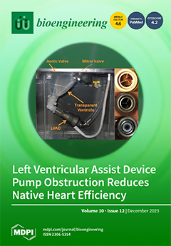
Obstruction of the LVAD flow path can occur when blood clots or tissue overgrowth form within the inflow cannula, pump body, or outflow graft, and it can lead to thrombus, embolism, and stroke. The impact of progressive pump inflow obstruction (PO) on the pressure and flow dynamics of the LVAD-supported heart was measured in a mock circulatory loop. Pressure and flow decreased with PO, shifting more of the flow through the aortic valve such that the total flow decreased by 6–11% and decreased the efficiency of the work of the native heart up to 60%. PO restricts diastolic flow through the LVAD, which reduces mitral inflow and decreases the strength and energy of the intraventricular vortices. The changes in flow architecture produced by PO include flow stasis and increased shear, which predispose the system to thromboembolic risk. View this paper
  • Issues are regarded as officially published after their release is announced to the table of contents alert mailing list.
  • You may sign up for e-mail alerts to receive table of contents of newly released issues.
  • PDF is the official format for papers published in both, html and pdf forms. To view the papers in pdf format, click on the "PDF Full-text" link, and use the free Adobe Reader to open them.
Order results
Result details
Section
Select all
Export citation of selected articles as:
17 pages, 5562 KiB  
Article
A Real-Time Control Method for Upper Limb Exoskeleton Based on Active Torque Prediction Model
Bioengineering 2023, 10(12), 1441; https://doi.org/10.3390/bioengineering10121441 - 18 Dec 2023
Viewed by 637
Abstract
Exoskeleton rehabilitation robots have been widely used in the rehabilitation treatment of stroke patients. Clinical studies confirmed that rehabilitation training with active movement intentions could improve the effectiveness of rehabilitation treatment significantly. This research proposes a real-time control method for an upper limb [...] Read more.
Exoskeleton rehabilitation robots have been widely used in the rehabilitation treatment of stroke patients. Clinical studies confirmed that rehabilitation training with active movement intentions could improve the effectiveness of rehabilitation treatment significantly. This research proposes a real-time control method for an upper limb exoskeleton based on the active torque prediction model. To fulfill the goal of individualized and precise rehabilitation, this method has an adjustable parameter assist ratio that can change the strength of the assist torque under the same conditions. In this study, upper limb muscles’ EMG signals and elbow angle were chosen as the sources of control signals. The active torque prediction model was then trained using a BP neural network after appropriately extracting features. The model exhibited good accuracy on PC and embedded systems, according to the experimental results. In the embedded system, the RMSE of this model was 0.1956 N·m and 94.98%. In addition, the proposed real-time control system also had an extremely low delay of only 40 ms, which would significantly increase the adaptability of human–computer interactions. Full article
(This article belongs to the Special Issue Robotics in Medical Engineering)
Show Figures

Figure 1

20 pages, 1372 KiB  
Review
The Autonomization Principle in Vascularized Flaps: An Alternative Strategy for Composite Tissue Scaffold In Vivo Revascularization
Bioengineering 2023, 10(12), 1440; https://doi.org/10.3390/bioengineering10121440 - 18 Dec 2023
Viewed by 599
Abstract
Autonomization is a physiological process allowing a flap to develop neo-vascularization from the reconstructed wound bed. This phenomenon has been used since the early application of flap surgeries but still remains poorly understood. Reconstructive strategies have greatly evolved since, and fasciocutaneous flaps have [...] Read more.
Autonomization is a physiological process allowing a flap to develop neo-vascularization from the reconstructed wound bed. This phenomenon has been used since the early application of flap surgeries but still remains poorly understood. Reconstructive strategies have greatly evolved since, and fasciocutaneous flaps have progressively replaced muscle-based reconstructions, ensuring better functional outcomes with great reliability. However, plastic surgeons still encounter challenges in complex cases where conventional flap reconstruction reaches its limitations. Furthermore, emerging bioengineering applications, such as decellularized scaffolds allowing a complex extracellular matrix to be repopulated with autologous cells, also face the complexity of revascularization. The objective of this article is to gather evidence of autonomization phenomena. A systematic review of flap autonomization is then performed to document the minimum delay allowing this process. Finally, past and potential applications in bio- and tissue-engineering approaches are discussed, highlighting the potential for in vivo revascularization of acellular scaffolds. Full article
Show Figures

Graphical abstract

13 pages, 535 KiB  
Article
Improved Hypertension Risk Assessment with Photoplethysmographic Recordings Combining Deep Learning and Calibration
Bioengineering 2023, 10(12), 1439; https://doi.org/10.3390/bioengineering10121439 - 18 Dec 2023
Viewed by 609
Abstract
Hypertension, a primary risk factor for various cardiovascular diseases, is a global health concern. Early identification and effective management of hypertensive individuals are vital for reducing associated health risks. This study explores the potential of deep learning (DL) techniques, specifically GoogLeNet, ResNet-18, and [...] Read more.
Hypertension, a primary risk factor for various cardiovascular diseases, is a global health concern. Early identification and effective management of hypertensive individuals are vital for reducing associated health risks. This study explores the potential of deep learning (DL) techniques, specifically GoogLeNet, ResNet-18, and ResNet-50, for discriminating between normotensive (NTS) and hypertensive (HTS) individuals using photoplethysmographic (PPG) recordings. The research assesses the impact of calibration at different time intervals between measurements, considering intervals less than 1 h, 1–6 h, 6–24 h, and over 24 h. Results indicate that calibration is most effective when measurements are closely spaced, with an accuracy exceeding 90% in all the DL strategies tested. For calibration intervals below 1 h, ResNet-18 achieved the highest accuracy (93.32%), sensitivity (84.09%), specificity (97.30%), and F1-score (88.36%). As the time interval between calibration and test measurements increased, classification performance gradually declined. For intervals exceeding 6 h, accuracy dropped below 81% but with all models maintaining accuracy above 71% even for intervals above 24 h. This study provides valuable insights into the feasibility of using DL for hypertension risk assessment, particularly through PPG recordings. It demonstrates that closely spaced calibration measurements can lead to highly accurate classification, emphasizing the potential for real-time applications. These findings may pave the way for advanced, non-invasive, and continuous blood pressure monitoring methods that are both efficient and reliable. Full article
(This article belongs to the Special Issue Machine Learning for Biomedical Applications, Volume II)
Show Figures

Figure 1

20 pages, 4185 KiB  
Article
Electroconductive Nanofibrous Scaffolds Enable Neuronal Differentiation in Response to Electrical Stimulation without Exogenous Inducing Factors
Bioengineering 2023, 10(12), 1438; https://doi.org/10.3390/bioengineering10121438 - 18 Dec 2023
Viewed by 714
Abstract
Among the various biochemical and biophysical inducers for neural regeneration, electrical stimulation (ES) has recently attracted considerable attention as an efficient means to induce neuronal differentiation in tissue engineering approaches. The aim of this in vitro study was to develop a nanofibrous scaffold [...] Read more.
Among the various biochemical and biophysical inducers for neural regeneration, electrical stimulation (ES) has recently attracted considerable attention as an efficient means to induce neuronal differentiation in tissue engineering approaches. The aim of this in vitro study was to develop a nanofibrous scaffold that enables ES-mediated neuronal differentiation in the absence of exogenous soluble inducers. A nanofibrous scaffold composed of polycaprolactone (PCL), poly-L-lactic acid (PLLA), and single-walled nanotubes (SWNTs) was fabricated via electrospinning and its physicochemical properties were investigated. The cytocompatibility of the electrospun composite with the PC12 cell line and bone marrow-derived mesenchymal stem cells (BMSCs) was investigated. The results showed that the PCL/PLLA/SWNT nanofibrous scaffold did not exhibit cytotoxicity and supported cell attachment, spreading, and proliferation. ES was applied to cells cultured on the nanofibrous scaffolds at different intensities and the expression of the three neural markers (Nestin, Microtubule-associated protein 2, and β tubulin-3) was evaluated using RT-qPCR analysis. The results showed that the highest expression of neural markers could be achieved at an electric field intensity of 200 mV/cm, suggesting that the scaffold in combination with ES can be an efficient tool to accelerate neural differentiation in the absence of exogenous soluble inducers. This has important implications for the regeneration of nerve injuries and may provide insights for further investigations of the mechanisms underlying ES-mediated neuronal commitment. Full article
Show Figures

Graphical abstract

12 pages, 3813 KiB  
Article
Exploring the Role of Desmoplastic Physical Stroma in Pancreatic Cancer Progression Using a Three-Dimensional Collagen Matrix Model
Bioengineering 2023, 10(12), 1437; https://doi.org/10.3390/bioengineering10121437 - 18 Dec 2023
Viewed by 848
Abstract
Pancreatic ductal adenocarcinoma (PDAC) is a refractory tumor with a poor prognosis, and its complex microenvironment is characterized by a fibrous interstitial matrix surrounding PDAC cells. Type I collagen is a major component of this interstitial matrix. Abundant type I collagen promotes its [...] Read more.
Pancreatic ductal adenocarcinoma (PDAC) is a refractory tumor with a poor prognosis, and its complex microenvironment is characterized by a fibrous interstitial matrix surrounding PDAC cells. Type I collagen is a major component of this interstitial matrix. Abundant type I collagen promotes its deposition and cross-linking to form a rigid and dense physical barrier, which limits drug penetration and immune cell infiltration and provides drug resistance and metabolic adaptations. In this study, to identify the physical effect of the stroma, type I collagen was used as a 3D matrix to culture Capan-1 cells and generate a 3D PDAC model. Using transcriptome analysis, a link between type I collagen-induced physical effects and the promotion of Capan-1 cell proliferation and migration was determined. Moreover, metabolomic analysis revealed that the physical effect caused a shift in metabolism toward a glycolytic phenotype. In particular, the high expression of proline in the metabolites suggests the ability to maintain Capan-1 cell proliferation under hypoxic and nutrient-depleted conditions. In conclusion, we identified type I collagen-induced physical effects in promoting Capan-1 cells, which cause PDAC progression, providing support for the role of dense stroma in the PDAC microenvironment and identifying a fundamental method for modeling the complex PDAC microenvironment. Full article
(This article belongs to the Special Issue The New Frontiers of Artificial Organs Engineering)
Show Figures

Figure 1

12 pages, 2304 KiB  
Article
A Virtual Inner Ear Model Selects Ramped Pulse Shapes for Vestibular Afferent Stimulation
Bioengineering 2023, 10(12), 1436; https://doi.org/10.3390/bioengineering10121436 - 18 Dec 2023
Viewed by 847
Abstract
Bilateral vestibular deficiency (BVD) results in chronic dizziness, blurry vision when moving the head, and postural instability. Vestibular prostheses (VPs) show promise as a treatment, but the VP-restored vestibulo-ocular reflex (VOR) gain in human trials falls short of expectations. We hypothesize that the [...] Read more.
Bilateral vestibular deficiency (BVD) results in chronic dizziness, blurry vision when moving the head, and postural instability. Vestibular prostheses (VPs) show promise as a treatment, but the VP-restored vestibulo-ocular reflex (VOR) gain in human trials falls short of expectations. We hypothesize that the slope of the rising ramp in stimulation pulses plays an important role in the recruitment of vestibular afferent units. To test this hypothesis, we utilized customized programming to generate ramped pulses with different slopes, testing their efficacy in inducing electrically evoked compound action potentials (eCAPs) and current spread via bench tests and simulations in a virtual inner model created in this study. The results confirmed that the slope of the ramping pulses influenced the recruitment of vestibular afferent units. Subsequently, an optimized stimulation pulse train was identified using model simulations, exhibiting improved modulation of vestibular afferent activity. This optimized slope not only reduced the excitation spread within the semicircular canals (SCCs) but also expanded the neural dynamic range. While the model simulations exhibited promising results, in vitro and in vivo experiments are warranted to validate the findings of this study in future investigations. Full article
(This article belongs to the Special Issue Engineering of Ears)
Show Figures

Figure 1

21 pages, 3292 KiB  
Review
How Artificial Intelligence Is Shaping Medical Imaging Technology: A Survey of Innovations and Applications
Bioengineering 2023, 10(12), 1435; https://doi.org/10.3390/bioengineering10121435 - 18 Dec 2023
Viewed by 1387
Abstract
The integration of artificial intelligence (AI) into medical imaging has guided in an era of transformation in healthcare. This literature review explores the latest innovations and applications of AI in the field, highlighting its profound impact on medical diagnosis and patient care. The [...] Read more.
The integration of artificial intelligence (AI) into medical imaging has guided in an era of transformation in healthcare. This literature review explores the latest innovations and applications of AI in the field, highlighting its profound impact on medical diagnosis and patient care. The innovation segment explores cutting-edge developments in AI, such as deep learning algorithms, convolutional neural networks, and generative adversarial networks, which have significantly improved the accuracy and efficiency of medical image analysis. These innovations have enabled rapid and accurate detection of abnormalities, from identifying tumors during radiological examinations to detecting early signs of eye disease in retinal images. The article also highlights various applications of AI in medical imaging, including radiology, pathology, cardiology, and more. AI-based diagnostic tools not only speed up the interpretation of complex images but also improve early detection of disease, ultimately delivering better outcomes for patients. Additionally, AI-based image processing facilitates personalized treatment plans, thereby optimizing healthcare delivery. This literature review highlights the paradigm shift that AI has brought to medical imaging, highlighting its role in revolutionizing diagnosis and patient care. By combining cutting-edge AI techniques and their practical applications, it is clear that AI will continue shaping the future of healthcare in profound and positive ways. Full article
(This article belongs to the Special Issue AI and Big Data Research in Biomedical Engineering)
Show Figures

Figure 1

12 pages, 4593 KiB  
Article
Modelling and Simulation of the Combined Use of IABP and Impella as a Rescue Procedure in Cardiogenic Shock: An Alternative for Non-Transplant Centres?
Bioengineering 2023, 10(12), 1434; https://doi.org/10.3390/bioengineering10121434 - 17 Dec 2023
Viewed by 588
Abstract
The treatment of critically ill patients remains an evolving and controversial issue. Mechanical circulatory support through a percutaneous approach for the management of cardiogenic shock has taken place in recent years. The combined use of IABP and the Impella 2.5 device may have [...] Read more.
The treatment of critically ill patients remains an evolving and controversial issue. Mechanical circulatory support through a percutaneous approach for the management of cardiogenic shock has taken place in recent years. The combined use of IABP and the Impella 2.5 device may have a role to play for this group of patients. A simulation approach may help with a quantitative assessment of the achievable degree of assistance. In this paper, we analyse the interaction between the Impella 2.5 pump applied as the LVAD and IABP using the numerical simulator of the cardiovascular system developed in our laboratory. Starting with pathological conditions reproduced using research data, the simulations were performed by setting different rotational speeds for the LVAD and driving the IABP in full mode (1:1) or partial mode (1:2, 1:3 and 1:4). The effects induced by drug administration during the assistance were also simulated. The haemodynamic parameters under investigation were aa follows: mean aortic pressure, systolic and diastolic aortic pressure, mean pulmonary arterial pressure, mean left and right atrial pressure, cardiac output, cardiac index, left and right ventricular end-systolic volume, left ventricular end-diastolic volume and mean coronary blood flow. The energetic variables considered in this study were as follows: left and right ventricular external work and left and right atrial pressure-volume area. The outcome of our simulations shows that the combined use of IABP and Impella 2.5 achieves adequate support in the acute phase of cardiogenic shock compared to each standalone device. This would allow further stabilisation and transfer to a transplant centre should the escalation of treatment be required. Full article
(This article belongs to the Special Issue Advances in Computational Modelling of Abdominal Aortic Aneurysm)
Show Figures

Figure 1

18 pages, 27457 KiB  
Article
Combined Edge Loss UNet for Optimized Segmentation in Total Knee Arthroplasty Preoperative Planning
Bioengineering 2023, 10(12), 1433; https://doi.org/10.3390/bioengineering10121433 - 16 Dec 2023
Viewed by 741
Abstract
Bone segmentation and 3D reconstruction are crucial for total knee arthroplasty (TKA) surgical planning with Personalized Surgical Instruments (PSIs). Traditional semi-automatic approaches are time-consuming and operator-dependent, although they provide reliable outcomes. Moreover, the recent expansion of artificial intelligence (AI) tools towards various medical [...] Read more.
Bone segmentation and 3D reconstruction are crucial for total knee arthroplasty (TKA) surgical planning with Personalized Surgical Instruments (PSIs). Traditional semi-automatic approaches are time-consuming and operator-dependent, although they provide reliable outcomes. Moreover, the recent expansion of artificial intelligence (AI) tools towards various medical domains is transforming modern healthcare. Accordingly, this study introduces an automated AI-based pipeline to replace the current operator-based tibia and femur 3D reconstruction procedure enhancing TKA preoperative planning. Leveraging an 822 CT image dataset, a novel patch-based method and an improved segmentation label generation algorithm were coupled to a Combined Edge Loss UNet (CEL-UNet), a novel CNN architecture featuring an additional decoding branch to boost the bone boundary segmentation. Root Mean Squared Errors and Hausdorff distances compared the predicted surfaces to the reference bones showing median and interquartile values of 0.26 (0.19–0.36) mm and 0.24 (0.18–0.32) mm, and of 1.06 (0.73–2.15) mm and 1.43 (0.82–2.86) mm for the tibia and femur, respectively, outperforming previous results of our group, state-of-the-art, and UNet models. A feasibility analysis for a PSI-based surgical plan revealed sub-millimetric distance errors and sub-angular alignment uncertainties in the PSI contact areas and the two cutting planes. Finally, operational environment testing underscored the pipeline’s efficiency. More than half of the processed cases complied with the PSI prototyping requirements, reducing the overall time from 35 min to 13.1 s, while the remaining ones underwent a manual refinement step to achieve such PSI requirements, performing the procedure four to eleven times faster than the manufacturer standards. To conclude, this research advocates the need for real-world applicability and optimization of AI solutions in orthopedic surgical practice. Full article
(This article belongs to the Special Issue Artificial Intelligence in Surgery)
Show Figures

Figure 1

15 pages, 5984 KiB  
Article
Non-Invasive Electroanatomical Mapping: A State-Space Approach for Myocardial Current Density Estimation
Bioengineering 2023, 10(12), 1432; https://doi.org/10.3390/bioengineering10121432 - 16 Dec 2023
Viewed by 573
Abstract
Electroanatomical mapping is a method for creating a model of the electrophysiology of the human heart. Medical professionals routinely locate and ablate the site of origin of cardiac arrhythmias with invasive catheterization. Non-invasive localization takes the form of electrocardiographic (ECG) or magnetocardiographic (MCG) [...] Read more.
Electroanatomical mapping is a method for creating a model of the electrophysiology of the human heart. Medical professionals routinely locate and ablate the site of origin of cardiac arrhythmias with invasive catheterization. Non-invasive localization takes the form of electrocardiographic (ECG) or magnetocardiographic (MCG) imaging, where the goal is to reconstruct the electrical activity of the human heart. Non-invasive alternatives to catheter electroanatomical mapping would reduce patients’ risks and open new venues for treatment planning and prevention. This work introduces a new system state-based method for estimating the electrical activity of the human heart from MCG measurements. Our model enables arbitrary propagation paths and velocities. A Kalman filter optimally estimates the current densities under the given measurements and model parameters. In an outer optimization loop, these model parameters are then optimized via gradient descent. This paper aims to establish the foundation for future research by providing a detailed mathematical explanation of the algorithm. We demonstrate the feasibility of our method through a simplified one-layer simulation. Our results show that the algorithm can learn the propagation paths from the magnetic measurements. A threshold-based segmentation into healthy and pathological tissue yields a DICE score of 0.84, a recall of 0.77, and a precision of 0.93. Full article
(This article belongs to the Section Biosignal Processing)
Show Figures

Figure 1

13 pages, 2667 KiB  
Article
Comparison of Traditional Impression and 3D Ear Scanning Techniques, Earmold Comfort, and Audiology Clinical Implications: A Pilot Study
Bioengineering 2023, 10(12), 1431; https://doi.org/10.3390/bioengineering10121431 - 16 Dec 2023
Viewed by 615
Abstract
This study investigated clinical aspects of the traditional ear impression and 3D ear scanning techniques. Adult earmold-users and non-users participated in this study. The earmold-users also participated in the earmold comfort comparison study by wearing earmolds from both techniques, one set a week [...] Read more.
This study investigated clinical aspects of the traditional ear impression and 3D ear scanning techniques. Adult earmold-users and non-users participated in this study. The earmold-users also participated in the earmold comfort comparison study by wearing earmolds from both techniques, one set a week according to a randomized sequence. Multiple clinical aspects of both techniques according to the participants and audiology professionals were recorded. Results revealed a preference for the 3D-scanning technique, which was perceived as more comfortable although both techniques were perceived as safe. Although the earmolds might have issues from both techniques, there was no significant difference in the perception of earmolds. Experience with the specific technique can affect the responses from the professionals. Compared to the traditional technique, 3D-scans had higher fixed but less variable costs and procedure times. A special clinical case was included and indicated that 3D-scans could be an option for specific patients. This study led to a better understanding of the two techniques clinically. With increasing involvement of new technology and more young professionals joining the profession of audiology, 3D ear scanning could be a viable consideration for audiology practices. Full article
(This article belongs to the Special Issue Engineering of Ears)
Show Figures

Figure 1

16 pages, 7370 KiB  
Article
Enhancing Skin Lesion Detection: A Multistage Multiclass Convolutional Neural Network-Based Framework
Bioengineering 2023, 10(12), 1430; https://doi.org/10.3390/bioengineering10121430 - 15 Dec 2023
Viewed by 816
Abstract
The early identification and treatment of various dermatological conditions depend on the detection of skin lesions. Due to advancements in computer-aided diagnosis and machine learning approaches, learning-based skin lesion analysis methods have attracted much interest recently. Employing the concept of transfer learning, this [...] Read more.
The early identification and treatment of various dermatological conditions depend on the detection of skin lesions. Due to advancements in computer-aided diagnosis and machine learning approaches, learning-based skin lesion analysis methods have attracted much interest recently. Employing the concept of transfer learning, this research proposes a deep convolutional neural network (CNN)-based multistage and multiclass framework to categorize seven types of skin lesions. In the first stage, a CNN model was developed to classify skin lesion images into two classes, namely benign and malignant. In the second stage, the model was then used with the transfer learning concept to further categorize benign lesions into five subcategories (melanocytic nevus, actinic keratosis, benign keratosis, dermatofibroma, and vascular) and malignant lesions into two subcategories (melanoma and basal cell carcinoma). The frozen weights of the CNN developed–trained with correlated images benefited the transfer learning using the same type of images for the subclassification of benign and malignant classes. The proposed multistage and multiclass technique improved the classification accuracy of the online ISIC2018 skin lesion dataset by up to 93.4% for benign and malignant class identification. Furthermore, a high accuracy of 96.2% was achieved for subclassification of both classes. Sensitivity, specificity, precision, and F1-score metrics further validated the effectiveness of the proposed multistage and multiclass framework. Compared to existing CNN models described in the literature, the proposed approach took less time to train and had a higher classification rate. Full article
Show Figures

Figure 1

10 pages, 1600 KiB  
Article
Post-Operative Delirium and Cognitive Dysfunction in Aged Patients Undergoing Cardiac Surgery: A Randomized Comparison between Two Blood Oxygenators
Bioengineering 2023, 10(12), 1429; https://doi.org/10.3390/bioengineering10121429 - 15 Dec 2023
Viewed by 728
Abstract
In elderly patients undergoing cardiac surgery, extracorporeal circulation affects the incidence of post-operative delirium and cognitive impairment with an impact on quality of life and mortality. In this study, a new oxygenator system (RemoweLL 2) was tested against a conventional system to assess [...] Read more.
In elderly patients undergoing cardiac surgery, extracorporeal circulation affects the incidence of post-operative delirium and cognitive impairment with an impact on quality of life and mortality. In this study, a new oxygenator system (RemoweLL 2) was tested against a conventional system to assess its efficacy in reducing the onset of postoperative delirium and cognitive dysfunction and the levels of serum inflammatory markers. A total of 154 patients (>65 y.o.) undergoing cardiopulmonary bypass (CPB) were enrolled and randomly assigned to oxygenator RemoweLL 2 (n = 81) or to gold standard device Inspire (n = 73) between September 2019 and March 2022. The aims of the study were to assess the incidence of delirium and the cognitive decline by neuropsychiatric tests and the MoCa test intra-hospital and at 6 months after CPB. Inflammation biomarkers in both groups were also evaluated. Before the CPB, the experimental groups were comparable for all variables. After CPB, the incidence of severe post-operative delirium showed a better trend (p = 0.093) in patients assigned to RemoweLL 2 (16.0%) versus Inspire (26.0%). Differences in enolase levels (p = 0.049), white blood cells (p = 0.006), and neutrophils (p = 0.003) in favor of RemoweLL 2 were also found. The use of novel and better construction technologies in CPB oxygenator devices results in measurable better neurocognitive and neurological outcomes in the elderly population undergoing CPB. Full article
(This article belongs to the Special Issue Medical Assistive Devices for Cardiovascular Diseases)
Show Figures

Graphical abstract

18 pages, 5195 KiB  
Article
DSE-NN: Deeply Supervised Efficient Neural Network for Real-Time Remote Photoplethysmography
Bioengineering 2023, 10(12), 1428; https://doi.org/10.3390/bioengineering10121428 - 15 Dec 2023
Viewed by 729
Abstract
Non-contact remote photoplethysmography can be used in a variety of medical and healthcare fields by measuring vital signs continuously and unobtrusively. Recently, end-to-end deep learning methods have been proposed to replace the existing handcrafted features. However, since the existing deep learning methods are [...] Read more.
Non-contact remote photoplethysmography can be used in a variety of medical and healthcare fields by measuring vital signs continuously and unobtrusively. Recently, end-to-end deep learning methods have been proposed to replace the existing handcrafted features. However, since the existing deep learning methods are known as black box models, the problem of interpretability has been raised, and the same problem exists in the remote photoplethysmography (rPPG) network. In this study, we propose a method to visualize temporal and spectral representations for hidden layers, deeply supervise the spectral representation of intermediate layers through the depth of networks and optimize it for a lightweight model. The optimized network improves performance and enables fast training and inference times. The proposed spectral deep supervision helps to achieve not only high performance but also fast convergence speed through the regularization of the intermediate layers. The effect of the proposed methods was confirmed through a thorough ablation study on public datasets. As a result, similar or outperforming results were obtained in comparison to state-of-the-art models. In particular, our model achieved an RMSE of 1 bpm on the PURE dataset, demonstrating its high accuracy. Moreover, it excelled on the V4V dataset with an impressive RMSE of 6.65 bpm, outperforming other methods. We observe that our model began converging from the very first epoch, a significant improvement over other models in terms of learning efficiency. Our approach is expected to be generally applicable to models that learn spectral domain information as well as to the applications of regression that require the representations of periodicity. Full article
(This article belongs to the Section Biosignal Processing)
Show Figures

Figure 1

11 pages, 1027 KiB  
Perspective
A Fermentation State Marker Rule Design Task in Metabolic Engineering
Bioengineering 2023, 10(12), 1427; https://doi.org/10.3390/bioengineering10121427 - 15 Dec 2023
Viewed by 626
Abstract
There are several ways in which mathematical modeling is used in fermentation control, but mechanistic mathematical genome-scale models of metabolism within the cell have not been applied or implemented so far. As part of the metabolic engineering task setting, we propose that metabolite [...] Read more.
There are several ways in which mathematical modeling is used in fermentation control, but mechanistic mathematical genome-scale models of metabolism within the cell have not been applied or implemented so far. As part of the metabolic engineering task setting, we propose that metabolite fluxes and/or biomass growth rate be used to search for a fermentation steady state marker rule. During fermentation, the bioreactor control system can automatically detect the desired steady state using a logical marker rule. The marker rule identification can be also integrated with the production growth coupling approach, as presented in this study. A design of strain with marker rule is demonstrated on genome scale metabolic model iML1515 of Escherichia coli MG1655 proposing two gene deletions enabling a measurable marker rule for succinate production using glucose as a substrate. The marker rule example at glucose consumption 10.0 is: IF (specific growth rate μ is above 0.060 h−1, AND CO2 production under 1.0, AND ethanol production above 5.5), THEN succinate production is within the range 8.2–10, where all metabolic fluxes units are mmol ∗ gDW−1 ∗ h−1. An objective function for application in metabolic engineering, including productivity features and rule detecting sensor set characterizing parameters, is proposed. Two-phase approach to implementing marker rules in the cultivation control system is presented to avoid the need for a modeler during production. Full article
(This article belongs to the Section Biochemical Engineering)
Show Figures

Graphical abstract

11 pages, 4874 KiB  
Article
Revolutionizing Patient Monitoring in Age-Related Macular Degeneration: A Comparative Study on the Necessity and Efficiency of the AMD VIEWER
Bioengineering 2023, 10(12), 1426; https://doi.org/10.3390/bioengineering10121426 - 15 Dec 2023
Viewed by 638
Abstract
(1) Background: Age-related Macular Degeneration (AMD) is a critical condition leading to blindness, necessitating lifelong clinic visits for management, albeit with existing challenges in monitoring its long-term progression. This study introduced and assessed an innovative tool, the AMD long-term Information Viewer (AMD VIEWER), [...] Read more.
(1) Background: Age-related Macular Degeneration (AMD) is a critical condition leading to blindness, necessitating lifelong clinic visits for management, albeit with existing challenges in monitoring its long-term progression. This study introduced and assessed an innovative tool, the AMD long-term Information Viewer (AMD VIEWER), designed to offer a comprehensive display of crucial medical data—including visual acuity, central retinal thickness, macular volume, vitreous injection treatment history, and Optical Coherent Tomography (OCT) images—across an individual eye’s entire treatment course. (2) Methods: By analyzing visit frequencies of patients with a history of invasive AMD treatment, a comparative examination between a Dropout group and an Active group underscored the clinical importance of regular visits, particularly highlighting better treatment outcomes and maintained visual acuity in the Active group. (3) Results: The efficiency of AMD VIEWER was proven by comparing it to manual data input by optometrists, showing significantly faster data display with no errors, unlike the time-consuming and error-prone manual entries. Furthermore, an elicited Net Promoter Score (NPS) of 70 from 10 ophthalmologists strongly endorsed AMD VIEWER’s practical utility. (4) Conclusions: This study underscores the importance of regular clinic visits for AMD patients. It suggests the AMD VIEWER as an effective tool for improving treatment data management and display. Full article
Show Figures

Figure 1

18 pages, 5759 KiB  
Review
Application and Technical Challenges in Design, Cloning, and Transfer of Large DNA
Bioengineering 2023, 10(12), 1425; https://doi.org/10.3390/bioengineering10121425 - 15 Dec 2023
Viewed by 646
Abstract
In the field of synthetic biology, rapid advancements in DNA assembly and editing have made it possible to manipulate large DNA, even entire genomes. These advancements have facilitated the introduction of long metabolic pathways, the creation of large-scale disease models, and the design [...] Read more.
In the field of synthetic biology, rapid advancements in DNA assembly and editing have made it possible to manipulate large DNA, even entire genomes. These advancements have facilitated the introduction of long metabolic pathways, the creation of large-scale disease models, and the design and assembly of synthetic mega-chromosomes. Generally, the introduction of large DNA in host cells encompasses three critical steps: design-cloning-transfer. This review provides a comprehensive overview of the three key steps involved in large DNA transfer to advance the field of synthetic genomics and large DNA engineering. Full article
(This article belongs to the Special Issue Applications of Genomic Technology in Disease Outcome Prediction)
Show Figures

Figure 1

17 pages, 18115 KiB  
Article
High Precision Cervical Precancerous Lesion Classification Method Based on ConvNeXt
Bioengineering 2023, 10(12), 1424; https://doi.org/10.3390/bioengineering10121424 - 15 Dec 2023
Viewed by 639
Abstract
Traditional cervical cancer diagnosis mainly relies on human papillomavirus (HPV) concentration testing. Considering that HPV concentrations vary from individual to individual and fluctuate over time, this method requires multiple tests, leading to high costs. Recently, some scholars have focused on the method of [...] Read more.
Traditional cervical cancer diagnosis mainly relies on human papillomavirus (HPV) concentration testing. Considering that HPV concentrations vary from individual to individual and fluctuate over time, this method requires multiple tests, leading to high costs. Recently, some scholars have focused on the method of cervical cytology for diagnosis. However, cervical cancer cells have complex textural characteristics and small differences between different cell subtypes, which brings great challenges for high-precision screening of cervical cancer. In this paper, we propose a high-precision cervical cancer precancerous lesion screening classification method based on ConvNeXt, utilizing self-supervised data augmentation and ensemble learning strategies to achieve cervical cancer cell feature extraction and inter-class discrimination, respectively. We used the Deep Cervical Cytological Levels (DCCL) dataset, which includes 1167 cervical cytology specimens from participants aged 32 to 67, for algorithm training and validation. We tested our method on the DCCL dataset, and the final classification accuracy was 8.85% higher than that of previous advanced models, which means that our method has significant advantages compared to other advanced methods. Full article
(This article belongs to the Special Issue Robotics in Medical Engineering)
Show Figures

Graphical abstract

15 pages, 3566 KiB  
Article
Application of a Radiomics Machine Learning Model for Differentiating Aldosterone-Producing Adenoma from Non-Functioning Adrenal Adenoma
Bioengineering 2023, 10(12), 1423; https://doi.org/10.3390/bioengineering10121423 - 14 Dec 2023
Viewed by 584
Abstract
To evaluate the secretory function of adrenal incidentaloma, this study explored the usefulness of a contrast-enhanced computed tomography (CECT)-based radiomics model for distinguishing aldosterone-producing adenoma (APA) from non-functioning adrenal adenoma (NAA). Overall, 68 APA and 60 NAA patients were randomly assigned (8:2 ratio) [...] Read more.
To evaluate the secretory function of adrenal incidentaloma, this study explored the usefulness of a contrast-enhanced computed tomography (CECT)-based radiomics model for distinguishing aldosterone-producing adenoma (APA) from non-functioning adrenal adenoma (NAA). Overall, 68 APA and 60 NAA patients were randomly assigned (8:2 ratio) to either a training or a test cohort. In the training cohort, univariate and least absolute shrinkage and selection operator regression analyses were conducted to select the significant features. A logistic regression machine learning (ML) model was then constructed based on the radiomics score and clinical features. Model effectiveness was evaluated according to the receiver operating characteristic, accuracy, sensitivity, specificity, F1 score, calibration plots, and decision curve analysis. In the test cohort, the area under the curve (AUC) of the Radscore model was 0.869 [95% confidence interval (CI), 0.734–1.000], and the accuracy, sensitivity, specificity, and F1 score were 0.731, 1.000, 0.583, and 0.900, respectively. The Clinic–Radscore model had an AUC of 0.994 [95% CI, 0.978–1.000], and the accuracy, sensitivity, specificity, and F1 score values were 0.962, 0.929, 1.000, and 0.931, respectively. In conclusion, the CECT-based radiomics and clinical radiomics ML model exhibited good diagnostic efficacy in differentiating APAs from NAAs; this non-invasive, cost-effective, and efficient method is important for the management of adrenal incidentaloma. Full article
Show Figures

Figure 1

21 pages, 5251 KiB  
Article
Anti-Apoptosis Therapy for Meniscal Avascular Zone Repair: A Proof-of-Concept Study in a Lapine Model
Bioengineering 2023, 10(12), 1422; https://doi.org/10.3390/bioengineering10121422 - 14 Dec 2023
Viewed by 550
Abstract
In the present study, 24 rabbits were firstly used to evaluate the apoptosis index and matrix degeneration after untreated adult meniscal tears. Vertical tears (0.25 cm in length) were prepared in the avascular zone of the anterior horn. Specimens were harvested at 1, [...] Read more.
In the present study, 24 rabbits were firstly used to evaluate the apoptosis index and matrix degeneration after untreated adult meniscal tears. Vertical tears (0.25 cm in length) were prepared in the avascular zone of the anterior horn. Specimens were harvested at 1, 3, 6, 12 weeks postoperatively. The apoptosis index around tear sites stayed at a high level throughout the whole follow-up period. The depletion of glycosaminoglycans (GAG) and aggrecan at the tear site was observed, while the deposition of COL I and COL II was not affected, even at the last follow-up of 12 weeks after operation. The expression of SOX9 decreased significantly; no cellularity was observed at the wound interface at all timepoints. Secondly, another 20 rabbits were included to evaluate the effects of anti-apoptosis therapy on rescuing meniscal cells and enhancing meniscus repair. Longitudinal vertical tears (0.5 cm in length) were made in the meniscal avascular body. Tears were repaired by the inside-out suture technique, or repaired with sutures in addition to fibrin gel and blank silica nanoparticles, or silica nanoparticles encapsulating apoptosis inhibitors (z-vad-fmk). Samples were harvested at 12 months postoperatively. We found the locally administered z-vad-fmk agent at the wound interface significantly alleviated meniscal cell apoptosis and matrix degradation, and enhanced meniscal repair in the avascular zone at 12 months after operation. Thus, local administration of caspase inhibitors (z-vad-fmk) is a promising therapeutic strategy for alleviating meniscal cell loss and enhancing meniscal repair after adult meniscal tears in the avascular zone. Full article
Show Figures

Figure 1

24 pages, 13599 KiB  
Article
A Critical Assessment of Generative Models for Synthetic Data Augmentation on Limited Pneumonia X-ray Data
Bioengineering 2023, 10(12), 1421; https://doi.org/10.3390/bioengineering10121421 - 14 Dec 2023
Viewed by 1158
Abstract
In medical imaging, deep learning models serve as invaluable tools for expediting diagnoses and aiding specialized medical professionals in making clinical decisions. However, effectively training deep learning models typically necessitates substantial quantities of high-quality data, a resource often lacking in numerous medical imaging [...] Read more.
In medical imaging, deep learning models serve as invaluable tools for expediting diagnoses and aiding specialized medical professionals in making clinical decisions. However, effectively training deep learning models typically necessitates substantial quantities of high-quality data, a resource often lacking in numerous medical imaging scenarios. One way to overcome this deficiency is to artificially generate such images. Therefore, in this comparative study we train five generative models to artificially increase the amount of available data in such a scenario. This synthetic data approach is evaluated on a a downstream classification task, predicting four causes for pneumonia as well as healthy cases on 1082 chest X-ray images. Quantitative and medical assessments show that a Generative Adversarial Network (GAN)-based approach significantly outperforms more recent diffusion-based approaches on this limited dataset with better image quality and pathological plausibility. We show that better image quality surprisingly does not translate to improved classification performance by evaluating five different classification models and varying the amount of additional training data. Class-specific metrics like precision, recall, and F1-score show a substantial improvement by using synthetic images, emphasizing the data rebalancing effect of less frequent classes. However, overall performance does not improve for most models and configurations, except for a DreamBooth approach which shows a +0.52 improvement in overall accuracy. The large variance of performance impact in this study suggests a careful consideration of utilizing generative models for limited data scenarios, especially with an unexpected negative correlation between image quality and downstream classification improvement. Full article
(This article belongs to the Special Issue Artificial Intelligence (AI) for Medical Image Processing)
Show Figures

Figure 1

22 pages, 7161 KiB  
Article
Revolutionizing Early Disease Detection: A High-Accuracy 4D CNN Model for Type 2 Diabetes Screening in Oman
Bioengineering 2023, 10(12), 1420; https://doi.org/10.3390/bioengineering10121420 - 14 Dec 2023
Viewed by 624
Abstract
The surge of diabetes poses a significant global health challenge, particularly in Oman and the Middle East. Early detection of diabetes is crucial for proactive intervention and improved patient outcomes. This research leverages the power of machine learning, specifically Convolutional Neural Networks (CNNs), [...] Read more.
The surge of diabetes poses a significant global health challenge, particularly in Oman and the Middle East. Early detection of diabetes is crucial for proactive intervention and improved patient outcomes. This research leverages the power of machine learning, specifically Convolutional Neural Networks (CNNs), to develop an innovative 4D CNN model dedicated to early diabetes prediction. A region-specific dataset from Oman is utilized to enhance health outcomes for individuals at risk of developing diabetes. The proposed model showcases remarkable accuracy, achieving an average accuracy of 98.49% to 99.17% across various epochs. Additionally, it demonstrates excellent F1 scores, recall, and sensitivity, highlighting its ability to identify true positive cases. The findings contribute to the ongoing effort to combat diabetes and pave the way for future research in using deep learning for early disease detection and proactive healthcare. Full article
(This article belongs to the Special Issue Recent Advances in Deep Learning: From Screening to Prognosis)
Show Figures

Graphical abstract

24 pages, 6029 KiB  
Article
Multi-Instance Classification of Breast Tumor Ultrasound Images Using Convolutional Neural Networks and Transfer Learning
Bioengineering 2023, 10(12), 1419; https://doi.org/10.3390/bioengineering10121419 - 13 Dec 2023
Viewed by 697
Abstract
Background: Breast cancer is arguably one of the leading causes of death among women around the world. The automation of the early detection process and classification of breast masses has been a prominent focus for researchers in the past decade. The utilization of [...] Read more.
Background: Breast cancer is arguably one of the leading causes of death among women around the world. The automation of the early detection process and classification of breast masses has been a prominent focus for researchers in the past decade. The utilization of ultrasound imaging is prevalent in the diagnostic evaluation of breast cancer, with its predictive accuracy being dependent on the expertise of the specialist. Therefore, there is an urgent need to create fast and reliable ultrasound image detection algorithms to address this issue. Methods: This paper aims to compare the efficiency of six state-of-the-art, fine-tuned deep learning models that can classify breast tissue from ultrasound images into three classes: benign, malignant, and normal, using transfer learning. Additionally, the architecture of a custom model is introduced and trained from the ground up on a public dataset containing 780 images, which was further augmented to 3900 and 7800 images, respectively. What is more, the custom model is further validated on another private dataset containing 163 ultrasound images divided into two classes: benign and malignant. The pre-trained architectures used in this work are ResNet-50, Inception-V3, Inception-ResNet-V2, MobileNet-V2, VGG-16, and DenseNet-121. The performance evaluation metrics that are used in this study are as follows: Precision, Recall, F1-Score and Specificity. Results: The experimental results show that the models trained on the augmented dataset with 7800 images obtained the best performance on the test set, having 94.95 ± 0.64%, 97.69 ± 0.52%, 97.69 ± 0.13%, 97.77 ± 0.29%, 95.07 ± 0.41%, 98.11 ± 0.10%, and 96.75 ± 0.26% accuracy for the ResNet-50, MobileNet-V2, InceptionResNet-V2, VGG-16, Inception-V3, DenseNet-121, and our model, respectively. Conclusion: Our proposed model obtains competitive results, outperforming some state-of-the-art models in terms of accuracy and training time. Full article
(This article belongs to the Special Issue Artificial Intelligence in Biomedical Imaging)
Show Figures

Figure 1

14 pages, 3088 KiB  
Article
Human Induced Pluripotent Spheroids’ Growth Is Driven by Viscoelastic Properties and Macrostructure of 3D Hydrogel Environment
Bioengineering 2023, 10(12), 1418; https://doi.org/10.3390/bioengineering10121418 - 13 Dec 2023
Viewed by 775
Abstract
Stem cells, particularly human iPSCs, constitute a powerful tool for tissue engineering, notably through spheroid and organoid models. While the sensitivity of stem cells to the viscoelastic properties of their direct microenvironment is well-described, stem cell differentiation still relies on biochemical factors. Our [...] Read more.
Stem cells, particularly human iPSCs, constitute a powerful tool for tissue engineering, notably through spheroid and organoid models. While the sensitivity of stem cells to the viscoelastic properties of their direct microenvironment is well-described, stem cell differentiation still relies on biochemical factors. Our aim is to investigate the role of the viscoelastic properties of hiPSC spheroids’ direct environment on their fate. To ensure that cell growth is driven only by mechanical interaction, bioprintable alginate–gelatin hydrogels with significantly different viscoelastic properties were utilized in differentiation factor-free culture medium. Alginate–gelatin hydrogels of varying concentrations were developed to provide 3D environments of significantly different mechanical properties, ranging from 1 to 100 kPa, while allowing printability. hiPSC spheroids from two different cell lines were prepared by aggregation (⌀ = 100 µm, n > 1 × 10⁴), included and cultured in the different hydrogels for 14 days. While spheroids within dense hydrogels exhibited limited growth, irrespective of formulation, porous hydrogels prepared with a liquid–liquid emulsion method displayed significant variations of spheroid morphology and growth as a function of hydrogel mechanical properties. Transversal culture (adjacent spheroids-laden alginate–gelatin hydrogels) clearly confirmed the separate effect of each hydrogel environment on hiPSC spheroid behavior. This study is the first to demonstrate that a mechanically modulated microenvironment induces diverse hiPSC spheroid behavior without the influence of other factors. It allows one to envision the combination of multiple formulations to create a complex object, where the fate of hiPSCs will be independently controlled by their direct microenvironment. Full article
(This article belongs to the Special Issue Advances in Hydrogels for Tissue Engineering Applications)
Show Figures

Graphical abstract

14 pages, 1281 KiB  
Article
Development and Validation of an Artificial Intelligence Preoperative Planning and Patient-Specific Instrumentation System for Total Knee Arthroplasty
Bioengineering 2023, 10(12), 1417; https://doi.org/10.3390/bioengineering10121417 - 13 Dec 2023
Viewed by 611
Abstract
Background: Accurate preoperative planning for total knee arthroplasty (TKA) is crucial. Computed tomography (CT)-based preoperative planning offers more comprehensive information and can also be used to design patient-specific instrumentation (PSI), but it requires well-reconstructed and segmented images, and the process is complex and [...] Read more.
Background: Accurate preoperative planning for total knee arthroplasty (TKA) is crucial. Computed tomography (CT)-based preoperative planning offers more comprehensive information and can also be used to design patient-specific instrumentation (PSI), but it requires well-reconstructed and segmented images, and the process is complex and time-consuming. This study aimed to develop an artificial intelligence (AI) preoperative planning and PSI system for TKA and to validate its time savings and accuracy in clinical applications. Methods: The 3D-UNet and modified HRNet neural network structures were used to develop the AI preoperative planning and PSI system (AIJOINT). Forty-two patients who were scheduled for TKA underwent both AI and manual CT processing and planning for component sizing, 20 of whom had their PSIs designed and applied intraoperatively. The time consumed and the size and orientation of the postoperative component were recorded. Results: The Dice similarity coefficient (DSC) and loss function indicated excellent performance of the neural network structure in CT image segmentation. AIJOINT was faster than conventional methods for CT segmentation (3.74 ± 0.82 vs. 128.88 ± 17.31 min, p < 0.05) and PSI design (35.10 ± 3.98 vs. 159.52 ± 17.14 min, p < 0.05) without increasing the time for size planning. The accuracy of AIJOINT in planning the size of both femoral and tibial components was 92.9%, while the accuracy of the conventional method in planning the size of the femoral and tibial components was 42.9% and 47.6%, respectively (p < 0.05). In addition, AI-based PSI improved the accuracy of the hip–knee–ankle angle and reduced postoperative blood loss (p < 0.05). Conclusion: AIJOINT significantly reduces the time needed for CT processing and PSI design without increasing the time for size planning, accurately predicts the component size, and improves the accuracy of lower limb alignment in TKA patients, providing a meaningful supplement to the application of AI in orthopaedics. Full article
(This article belongs to the Special Issue Advanced Engineering Technology in Orthopaedic Research)
Show Figures

Figure 1

17 pages, 12907 KiB  
Article
High-Speed and Accurate Diagnosis of Gastrointestinal Disease: Learning on Endoscopy Images Using Lightweight Transformer with Local Feature Attention
Bioengineering 2023, 10(12), 1416; https://doi.org/10.3390/bioengineering10121416 - 13 Dec 2023
Viewed by 662
Abstract
In response to the pressing need for robust disease diagnosis from gastrointestinal tract (GIT) endoscopic images, we proposed FLATer, a fast, lightweight, and highly accurate transformer-based model. FLATer consists of a residual block, a vision transformer module, and a spatial attention block, which [...] Read more.
In response to the pressing need for robust disease diagnosis from gastrointestinal tract (GIT) endoscopic images, we proposed FLATer, a fast, lightweight, and highly accurate transformer-based model. FLATer consists of a residual block, a vision transformer module, and a spatial attention block, which concurrently focuses on local features and global attention. It can leverage the capabilities of both convolutional neural networks (CNNs) and vision transformers (ViT). We decomposed the classification of endoscopic images into two subtasks: a binary classification to discern between normal and pathological images and a further multi-class classification to categorize images into specific diseases, namely ulcerative colitis, polyps, and esophagitis. FLATer has exhibited exceptional prowess in these tasks, achieving 96.4% accuracy in binary classification and 99.7% accuracy in ternary classification, surpassing most existing models. Notably, FLATer could maintain impressive performance when trained from scratch, underscoring its robustness. In addition to the high precision, FLATer boasted remarkable efficiency, reaching a notable throughput of 16.4k images per second, which positions FLATer as a compelling candidate for rapid disease identification in clinical practice. Full article
(This article belongs to the Special Issue Recent Advance of Machine Learning in Biomedical Image Analysis)
Show Figures

Figure 1

16 pages, 4287 KiB  
Article
Towards Optimizing Sub-Normothermic Machine Perfusion in Fasciocutaneous Flaps: A Large Animal Study
Bioengineering 2023, 10(12), 1415; https://doi.org/10.3390/bioengineering10121415 - 12 Dec 2023
Cited by 1 | Viewed by 717
Abstract
Machine perfusion has developed rapidly since its first use in solid organ transplantation. Likewise, reconstructive surgery has kept pace, and ex vivo perfusion appears as a new trend in vascularized composite allotransplants preservation. In autologous reconstruction, fasciocutaneous flaps are now the gold standard [...] Read more.
Machine perfusion has developed rapidly since its first use in solid organ transplantation. Likewise, reconstructive surgery has kept pace, and ex vivo perfusion appears as a new trend in vascularized composite allotransplants preservation. In autologous reconstruction, fasciocutaneous flaps are now the gold standard due to their low morbidity (muscle sparing) and favorable functional and cosmetic results. However, failures still occasionally arise due to difficulties encountered with the vessels during free flap transfer. The development of machine perfusion procedures would make it possible to temporarily substitute or even avoid microsurgical anastomoses in certain complex cases. We performed oxygenated acellular sub-normothermic perfusions of fasciocutaneous flaps for 24 and 48 h in a porcine model and compared continuous and intermittent perfusion regimens. The monitored metrics included vascular resistance, edema, arteriovenous oxygen gas differentials, and metabolic parameters. A final histological assessment was performed. Porcine flaps which underwent successful oxygenated perfusion showed minimal or no signs of cell necrosis at the end of the perfusion. Intermittent perfusion allowed overall better results to be obtained at 24 h and extended perfusion duration. This work provides a strong foundation for further research and could lead to new and reliable reconstructive techniques. Full article
(This article belongs to the Section Biomedical Engineering and Biomaterials)
Show Figures

Graphical abstract

18 pages, 3753 KiB  
Article
Vibro-Acoustic Platelet Activation: An Additive Mechanism of Prothrombosis with Applicability to Snoring and Obstructive Sleep Apnea
Bioengineering 2023, 10(12), 1414; https://doi.org/10.3390/bioengineering10121414 - 12 Dec 2023
Viewed by 730
Abstract
Introduction: Obstructive sleep apnea (OSA) and loud snoring are conditions with increased cardiovascular risk and notably an association with stroke. Central in stroke are thrombosis and thromboembolism, all related to and initiaing with platelet activation. Platelet activation in OSA has been felt to [...] Read more.
Introduction: Obstructive sleep apnea (OSA) and loud snoring are conditions with increased cardiovascular risk and notably an association with stroke. Central in stroke are thrombosis and thromboembolism, all related to and initiaing with platelet activation. Platelet activation in OSA has been felt to be driven by biochemical and inflammatory means, including intermittent catecholamine exposure and transient hypoxia. We hypothesized that snore-associated acoustic vibration (SAAV) is an activator of platelets that synergizes with catecholamines and hypoxia to further amplify platelet activation. Methods: Gel-filtered human platelets were exposed to snoring utilizing a designed vibro-acoustic exposure device, varying the time and intensity of exposure and frequency content. Platelet activation was assessed via thrombin generation using the Platelet Activity State assay and scanning electron microscopy. Comparative activation induced by epinephrine and hypoxia were assessed individually as well as additively with SAAV, as well as the inhibitory effect of aspirin. Results: We demonstrate that snore-associated acoustic vibration is an independent activator of platelets, which is dependent upon the dose of exposure, i.e., intensity x time. In snoring, acoustic vibrations associated with low-frequency sound content (200 Hz) are more activating than those associated with high frequencies (900 Hz) (53.05% vs. 22.08%, p = 0.001). Furthermore, SAAV is additive to both catecholamines and hypoxia-mediated activation, inducing synergistic activation. Finally, aspirin, a known inhibitor of platelet activation, has no significant effect in limiting SAAV platelet activation. Conclusion: Snore-associated acoustic vibration is a mechanical means of platelet activation, which may drive prothrombosis and thrombotic risk clinically observed in loud snoring and OSA. Full article
Show Figures

Figure 1

13 pages, 2369 KiB  
Article
Biomechanical and Biological Assessment of Polyglycelrolsebacate-Coupled Implant with Shape Memory Effect for Treating Osteoporotic Fractures
Bioengineering 2023, 10(12), 1413; https://doi.org/10.3390/bioengineering10121413 - 12 Dec 2023
Viewed by 625
Abstract
Poly(glycerol sebacate) is a biocompatible elastomer that has gained increasing attention as a potential biomaterial for tissue engineering applications. In particular, PGS is capable of providing shape memory effects and allows for a free form, which can remember the original shape and obtain [...] Read more.
Poly(glycerol sebacate) is a biocompatible elastomer that has gained increasing attention as a potential biomaterial for tissue engineering applications. In particular, PGS is capable of providing shape memory effects and allows for a free form, which can remember the original shape and obtain a temporary shape under melting point and then can recover its original shape at body temperature. Because these properties can easily produce customized shapes, PGS is being coupled with implants to offer improved fixation and maintenance of implants for fractures of osteoporosis bone. Herein, this study fabricated the OP implant with a PGS membrane and investigated the potential of this coupling. Material properties were characterized and compared with various PGS membranes to assess features such as control of curing temperature, curing time, and washing time. Based on the ISO 10993-5 standard, in vitro cell culture studies with C2C12 cells confirmed that the OP implant coupled with PGS membrane showed biocompatibility and biomechanical experiments indicated significantly increased pullout strength and maintenance. It is believed that this multifunctional OP implant will be useful for bone tissue engineering applications. Full article
(This article belongs to the Special Issue Advanced Engineering Technology in Orthopaedic Research)
Show Figures

Figure 1

28 pages, 3359 KiB  
Article
Tuning Fatty Acid Profile and Yield in Pichia pastoris
Bioengineering 2023, 10(12), 1412; https://doi.org/10.3390/bioengineering10121412 - 12 Dec 2023
Viewed by 730
Abstract
Fatty acids have been supplied for diverse non-food, industrial applications from plant oils and animal fats for many decades. Due to the massively increasing world population demanding a nutritious diet and the thrive to provide feedstocks for industrial production lines in a sustainable [...] Read more.
Fatty acids have been supplied for diverse non-food, industrial applications from plant oils and animal fats for many decades. Due to the massively increasing world population demanding a nutritious diet and the thrive to provide feedstocks for industrial production lines in a sustainable way, i.e., independent from food supply chains, alternative fatty acid sources have massively gained in importance. Carbohydrate-rich side-streams of agricultural production, e.g., molasses, lignocellulosic waste, glycerol from biodiesel production, and even CO2, are considered and employed as carbon sources for the fermentative accumulation of fatty acids in selected microbial hosts. While certain fatty acid species are readily accumulated in native microbial metabolic routes, other fatty acid species are scarce, and host strains need to be metabolically engineered for their high-level production. We report the metabolic engineering of Pichia pastoris to produce palmitoleic acid from glucose and discuss the beneficial and detrimental engineering steps in detail. Fatty acid secretion was achieved through the deletion of fatty acyl-CoA synthetases and overexpression of the truncated E. coli thioesterase ‘TesA. The best strains secreted >1 g/L free fatty acids into the culture medium. Additionally, the introduction of C16-specific ∆9-desaturases and fatty acid synthases, coupled with improved cultivation conditions, increased the palmitoleic acid content from 5.5% to 22%. Full article
(This article belongs to the Section Biochemical Engineering)
Show Figures

Graphical abstract

Previous Issue
Next Issue
Back to TopTop