Next Issue
Volume 46, January
Previous Issue
Volume 45, November
 
 
cimb-logo

Journal Browser

Journal Browser

Curr. Issues Mol. Biol., Volume 45, Issue 12 (December 2023) – 54 articles

Cover Story (view full-size image): 
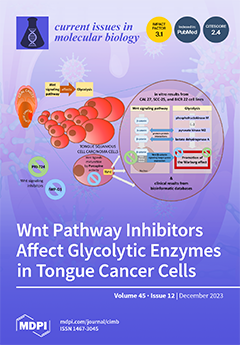
The dysregulation of energetic metabolism is one of the hallmarks of cancer cells. Indeed, the growth of head and neck cancer cells depends heavily on glycolytic activity, which can be considered a potential therapeutic target. Wnt signaling is one of the pathways that undergoes upregulation in HNSCC. We have shown that Wnt signaling inhibitors—PRI-724 and IWP-O1—affect glycolysis by attenuating the expression of key glycolytic genes—phosphofructokinase M, pyruvate kinase M2, and lactate dehydrogenase A, whose expression is increased in head and neck cancers, especially in HPV-positive cases. Thus, we provide evidence that inhibiting glucose metabolism via Wnt signaling inhibitors is a promising action against tongue cancer cells. View this paper
  • Issues are regarded as officially published after their release is announced to the table of contents alert mailing list.
  • You may sign up for e-mail alerts to receive table of contents of newly released issues.
  • PDF is the official format for papers published in both, html and pdf forms. To view the papers in pdf format, click on the "PDF Full-text" link, and use the free Adobe Reader to open them.
Order results
Result details
Section
Select all
Export citation of selected articles as:
24 pages, 14551 KiB  
Article
In Silico Analysis of SARS-CoV-2 Non-Structural Proteins Reveals an Interaction with the Host’s Heat Shock Proteins That May Contribute to Viral Replications and Development
Curr. Issues Mol. Biol. 2023, 45(12), 10225-10247; https://doi.org/10.3390/cimb45120638 - 18 Dec 2023
Viewed by 582
Abstract
The non-structural protein 2 (NSP2) is an RNA-binding protein involved in coronavirus genome replication, and it often decreases human immune response to promote viral invasion and development. It is believed that the NSP2 associates itself with polyamines and heat shock proteins inside the [...] Read more.
The non-structural protein 2 (NSP2) is an RNA-binding protein involved in coronavirus genome replication, and it often decreases human immune response to promote viral invasion and development. It is believed that the NSP2 associates itself with polyamines and heat shock proteins inside the host cell to proceed with viral development. This study aimed to investigate how the SARS-CoV-2 virus’ key non-structural proteins (NSP2) utilize polyamines and heat shock proteins using a molecular docking approach and molecular dynamics (MD). ClusPro and HADDOCK servers were used for the docking and Discovery Studio, chimera, and PyMOL were used for analysis. Docking of the heat shock proteins 40 (HSP40), 70 (HSP70), and 90 (HSP90) with SARS-CoV-2 NSP2 resulted in 32, 28, and 19 interactions, respectively. Molecular dynamics revealed Arg458, Asn508, Met297, Arg301, and Trp417 as active residues, and pharmacophore modeling indicated ZINC395648, ZINC01150525, and ZINC85324008 from the zinc database as possible inhibitors for this NSP2. Full article
(This article belongs to the Special Issue Predicting Drug Targets Using Bioinformatics Methods)
Show Figures

Figure 1

14 pages, 2629 KiB  
Article
Water Extract of Portulaca Oleracea Inhibits PEDV Infection-Induced Pyrolysis by Caspase-1/GSDMD
Curr. Issues Mol. Biol. 2023, 45(12), 10211-10224; https://doi.org/10.3390/cimb45120637 - 18 Dec 2023
Viewed by 583
Abstract
Porcine epidemic diarrhea virus (PEDV) belongs to the coronavirus family and the coronavirus genus, causing contact enteric infection in pigs. It is one of the most serious diseases that threatens the pig industry. However, there is currently no specific drug to prevent and [...] Read more.
Porcine epidemic diarrhea virus (PEDV) belongs to the coronavirus family and the coronavirus genus, causing contact enteric infection in pigs. It is one of the most serious diseases that threatens the pig industry. However, there is currently no specific drug to prevent and treat the disease, indicating that we need to be vigilant about the spread of the disease and the development of anti-PEDV drugs. The dried aerial parts of the plant Portulaca oleracea in the family Portulacaceous, whose decoction can be used to treat acute enteritis, dysentery, diarrhea, and other diseases. This study explored the potential mechanism of water extract of Portulaca oleracea (WEPO) in PEDV-induced pyroptosis in Vero cells. PEDV decreased the viability of Vero cells in a dose- and time-dependent manner, causing cell damage, upregulating the level of intracellular Nlrp3, and inhibiting the level of Gasdermin D (GSDMD) and the activation of Caspase-1. WEPO can inhibit PEDV-induced pyroptosis, reduce the elevation of inflammatory factors caused by infection, and exhibit a dose-dependent effect. Knockdown of Caspase-1 and GSDMD separately can induce the production of the inflammatory factor IL-1β to significantly decrease and increase, respectively. These results suggest that WEPO can inhibit cell pyroptosis caused by PEDV and that the Caspase-1 and GSDMD pathways play an important role in this process. Full article
Show Figures

Figure 1

18 pages, 10885 KiB  
Article
Hydrogen Attenuates Chronic Intermittent Hypoxia-Induced Cardiac Hypertrophy by Regulating Iron Metabolism
Curr. Issues Mol. Biol. 2023, 45(12), 10193-10210; https://doi.org/10.3390/cimb45120636 - 16 Dec 2023
Viewed by 465
Abstract
The present study aimed to investigate the impact of hydrogen (H2) on chronic intermittent hypoxia (CIH)-induced cardiac hypertrophy in mice by modulating iron metabolism. C57BL/6N mice were randomly allocated into four groups: control (Con), CIH, CIH + H2, and [...] Read more.
The present study aimed to investigate the impact of hydrogen (H2) on chronic intermittent hypoxia (CIH)-induced cardiac hypertrophy in mice by modulating iron metabolism. C57BL/6N mice were randomly allocated into four groups: control (Con), CIH, CIH + H2, and H2. The mice were exposed to CIH (21–5% FiO2, 3 min/cycle, 8 h/d), and received inhalation of a hydrogen–oxygen mixture (2 h/d) for 5 weeks. Cardiac and mitochondrial function, levels of reactive oxygen species (ROS), and iron levels were evaluated. The H9C2 cell line was subjected to intermittent hypoxia (IH) and treated with H2. Firstly, we found H2 had a notable impact on cardiac hypertrophy, ameliorated pathological alterations and mitochondrial morphology induced by CIH (p < 0.05). Secondly, H2 exhibited a suppressive effect on oxidative injury by decreasing levels of inducible nitric oxide synthase (i-NOS) (p < 0.05) and 4-hydroxynonenal (4-HNE) (p < 0.01). Thirdly, H2 demonstrated a significant reduction in iron levels within myocardial cells through the upregulation of ferroportin 1 (FPN1) proteins (p < 0.01) and the downregulation of transferrin receptor 1 (TfR1), divalent metal transporter 1 with iron-responsive element (DMT1(+ire)), and ferritin light chain (FTL) mRNA or proteins (p < 0.05). Simultaneously, H2 exhibited the ability to decrease the levels of Fe2+ and ROS in H9C2 cells exposed to IH (p < 0.05). Moreover, H2 mediated the expression of hepcidin, hypoxia-inducible factor-1α (HIF-1α) (p < 0.01), and iron regulatory proteins (IRPs), which might be involved in the regulation of iron-related transporter proteins. These results suggested that H2 may be beneficial in preventing cardiac hypertrophy, a condition associated with reduced iron toxicity. Full article
(This article belongs to the Special Issue Iron Metabolism: From Molecular Mechanisms to Molecular Imaging)
Show Figures

Figure 1

14 pages, 2785 KiB  
Article
Expression of Human L-Dopa Decarboxylase (DDC) under Conditions of Oxidative Stress
Curr. Issues Mol. Biol. 2023, 45(12), 10179-10192; https://doi.org/10.3390/cimb45120635 - 16 Dec 2023
Viewed by 363
Abstract
Oxidative stress is known to influence mRNA levels, translation, and proteolysis. The importance of oxidative stress has been demonstrated in several human diseases, including neurodegenerative disorders. L-Dopa decarboxylase (DDC) is the enzyme that converts L-Dopa to dopamine (DA). In spite of a large [...] Read more.
Oxidative stress is known to influence mRNA levels, translation, and proteolysis. The importance of oxidative stress has been demonstrated in several human diseases, including neurodegenerative disorders. L-Dopa decarboxylase (DDC) is the enzyme that converts L-Dopa to dopamine (DA). In spite of a large number of studies, little is known about the biological significance of the enzyme under physiological and pathological conditions. Here, we investigated the relationship between DDC expression and oxidative stress in human neural and non-neural cells. Oxidative stress was induced by treatment with H2O2. Our data indicated that mRNA and protein expression of DDC was enhanced or remained stable under conditions of ROS induction, despite degradation of total RNA and increased cytotoxicity and apoptosis. Moreover, DDC silencing caused an increase in the H2O2-induced cytotoxicity. The current study suggests that DDC is involved in the mechanisms of oxidative stress. Full article
(This article belongs to the Section Biochemistry, Molecular and Cellular Biology)
Show Figures

Figure 1

20 pages, 5486 KiB  
Article
Improvement in Yield of Extracellular Vesicles Derived from Edelweiss Callus Treated with LED Light and Enhancement of Skin Anti-Aging Indicators
Curr. Issues Mol. Biol. 2023, 45(12), 10159-10178; https://doi.org/10.3390/cimb45120634 - 16 Dec 2023
Viewed by 725
Abstract
The process of skin aging is currently recognized as a disease, and extracellular vesicles (EVs) are being used to care for it. While various EVs are present in the market, there is a growing need for research on improving skin conditions through microbial [...] Read more.
The process of skin aging is currently recognized as a disease, and extracellular vesicles (EVs) are being used to care for it. While various EVs are present in the market, there is a growing need for research on improving skin conditions through microbial and plant-derived EVs. Edelweiss is a medicinal plant and is currently an endangered species. Callus culture is a method used to protect rare medicinal plants, and recently, research on EVs using callus culture has been underway. In this study, the researchers used LED light to increase the productivity of Edelweiss EVs and confirmed that productivity was enhanced by LED exposure. Additionally, improvements in skin anti-aging indicators were observed. Notably, M-LED significantly elevated callus fresh and dry weight, with a DW/FW ratio of 4.11%, indicating enhanced proliferation. Furthermore, M-LED boosted secondary metabolite production, including a 20% increase in total flavonoids and phenolics. The study explores the influence of M-LED on EV production, revealing a 2.6-fold increase in concentration compared to darkness. This effect is consistent across different plant species (Centella asiatica, Panax ginseng), demonstrating the universality of the phenomenon. M-LED-treated EVs exhibit a concentration-dependent inhibition of reactive oxygen species (ROS) production, surpassing dark-cultured EVs. Extracellular melanin content analysis reveals M-LED-cultured EVs’ efficacy in reducing melanin production. Additionally, the expression of key skin proteins (FLG, AQP3, COL1) is significantly higher in fibroblasts treated with M-LED-cultured EVs. These results are expected to provide valuable insights into research on improving the productivity of plant-derived EVs and enhancing skin treatment using plant-derived EVs. Full article
(This article belongs to the Section Biochemistry, Molecular and Cellular Biology)
Show Figures

Figure 1

28 pages, 12633 KiB  
Review
Clinical Significance of SOX10 Expression in Human Pathology
Curr. Issues Mol. Biol. 2023, 45(12), 10131-10158; https://doi.org/10.3390/cimb45120633 - 15 Dec 2023
Viewed by 580
Abstract
The embryonic development of neural crest cells and subsequent tissue differentiation are intricately regulated by specific transcription factors. Among these, SOX10, a member of the SOX gene family, stands out. Located on chromosome 22q13, the SOX10 gene encodes a transcription factor crucial [...] Read more.
The embryonic development of neural crest cells and subsequent tissue differentiation are intricately regulated by specific transcription factors. Among these, SOX10, a member of the SOX gene family, stands out. Located on chromosome 22q13, the SOX10 gene encodes a transcription factor crucial for the differentiation, migration, and maintenance of tissues derived from neural crest cells. It plays a pivotal role in developing various tissues, including the central and peripheral nervous systems, melanocytes, chondrocytes, and odontoblasts. Mutations in SOX10 have been associated with congenital disorders such as Waardenburg–Shah Syndrome, PCWH syndrome, and Kallman syndrome, underscoring its clinical significance. Furthermore, SOX10 is implicated in neural and neuroectodermal tumors, such as melanoma, malignant peripheral nerve sheath tumors (MPNSTs), and schwannomas, influencing processes like proliferation, migration, and differentiation. In mesenchymal tumors, SOX10 expression serves as a valuable marker for distinguishing between different tumor types. Additionally, SOX10 has been identified in various epithelial neoplasms, including breast, ovarian, salivary gland, nasopharyngeal, and bladder cancers, presenting itself as a potential diagnostic and prognostic marker. However, despite these associations, further research is imperative to elucidate its precise role in these malignancies. Full article
(This article belongs to the Special Issue Advanced Molecular Solutions for Cancer Therapy)
Show Figures

Figure 1

10 pages, 12676 KiB  
Communication
NRG1 Regulates Proliferation, Migration and Differentiation of Human Limbal Epithelial Stem Cells
Curr. Issues Mol. Biol. 2023, 45(12), 10121-10130; https://doi.org/10.3390/cimb45120632 - 14 Dec 2023
Viewed by 483
Abstract
Limbal epithelial stem/progenitor cells (LESCs) proliferate, migrate and differentiate into mature corneal epithelium cells (CECs) that cover the ocular surface. LESCs play a crucial role in the maintenance and regeneration of the corneal epithelium, and their dysfunction can lead to various corneal diseases. [...] Read more.
Limbal epithelial stem/progenitor cells (LESCs) proliferate, migrate and differentiate into mature corneal epithelium cells (CECs) that cover the ocular surface. LESCs play a crucial role in the maintenance and regeneration of the corneal epithelium, and their dysfunction can lead to various corneal diseases. Neuregulin 1 (NRG1) is a member of the epidermal growth factor family that regulates the growth and differentiation of epithelial tissues. Here, we depicted the dynamic transcriptomic profiles during human CEC differentiation, identifying six gene co-expression modules that were specific to different differentiation stages. We found that the expression of NRG1 was high in human LESCs and decreased dramatically upon differentiation. Knockdown of NRG1 significantly inhibited LESC proliferation and upregulated the expression of the terminal differentiation marker genes KRT3, KRT12 and CLU. In addition, the scratch wound closure assay showed that knockdown of NRG1 attenuated wound closure of LESCs over 24 h. Together, we dissected the transcriptional regulatory dynamics during CEC differentiation and identified NRG1 as a key regulator that promoted LESC proliferation and migration and maintained the undifferentiated state. Full article
(This article belongs to the Special Issue Molecular Research in Stem Cells)
Show Figures

Figure 1

12 pages, 1228 KiB  
Article
The Cytotoxicity of Cotyledon orbiculata Aqueous Extract and the Biogenic Silver Nanoparticles Derived from the Extract
Curr. Issues Mol. Biol. 2023, 45(12), 10109-10120; https://doi.org/10.3390/cimb45120631 - 14 Dec 2023
Viewed by 511
Abstract
Green synthesized silver nanoparticles (AgNPs) have become popular because of their promising biological activities. However, for most of these nanoparticles, the cytotoxic effects have not been determined and their safety is not guaranteed. In a previous study, we successfully synthesized AgNPs (Cotyledon [...] Read more.
Green synthesized silver nanoparticles (AgNPs) have become popular because of their promising biological activities. However, for most of these nanoparticles, the cytotoxic effects have not been determined and their safety is not guaranteed. In a previous study, we successfully synthesized AgNPs (Cotyledon-AgNPs) using an extract of Cotyledon orbiculata, a medicinal plant traditionally used in South Africa to treat skin conditions. Cotyledon-AgNPs were shown to have significant antimicrobial and wound-healing activities. Fibroblast cells treated with extracts of C. orbiculata and Cotyledon-AgNPs demonstrated an enhanced growth rate, which is essential in wound healing. These nanoparticles therefore have promising wound-healing activities. However, the cytotoxicity of these nanoparticles is not known. In this study, the toxic effects of C. orbiculata extract and Cotyledon-AgNPs on the non-cancerous skin fibroblast (KMST-6) were determined using in vitro assays to assess oxidative stress and cell death. Both the C. orbiculata extract and the Cotyledon-AgNPs did not show any significant cytotoxic effects in these assays. Gene expression analysis was also used to assess the cytotoxic effects of Cotyledon-AgNPs at a molecular level. Of the eighty-four molecular toxicity genes analysed, only eight (FASN, SREBF1, CPT2, ASB1, HSPA1B, ABCC2, CASP9, and MKI67) were differentially expressed. These genes are mainly involved in fatty acid and mitochondrial energy metabolism. The results support the finding that Cotyledon-AgNPs have low cytotoxicity at the concentrations tested. The upregulation of genes such as FASN, SERBF1, and MKI-67 also support previous findings that Cotyledon-AgNPs can promote wound healing via cell growth and proliferation. It can therefore be concluded that Cotyledon-AgNPs are not toxic to skin fibroblast cells at the concentration that promotes wound healing. These nanoparticles could possibly be safely used for wound healing. Full article
(This article belongs to the Special Issue Effects of Nanoparticles on Living Organisms 2.0)
Show Figures

Figure 1

12 pages, 3198 KiB  
Article
Systemic Immune Modulation Induced by Ephedrine in Obese-Diabetes (db/db) Mice
Curr. Issues Mol. Biol. 2023, 45(12), 10097-10108; https://doi.org/10.3390/cimb45120630 - 14 Dec 2023
Viewed by 521
Abstract
Immune-modulatory effects in obese-diabetes (db/db) mice were observed to understand the possible mechanism(s) of ephedrine-induced unfavorable responses. The ephedrine doses were selected based on the FDA report (NTP Tech Rep Ser NO 307; CAS# 134-72-5), which showed the non-toxic dose for [...] Read more.
Immune-modulatory effects in obese-diabetes (db/db) mice were observed to understand the possible mechanism(s) of ephedrine-induced unfavorable responses. The ephedrine doses were selected based on the FDA report (NTP Tech Rep Ser NO 307; CAS# 134-72-5), which showed the non-toxic dose for B6C3F1 mice. In db/db mice, higher doses (6 and 12 mg/mouse) of ephedrine significantly harmed the liver and lung morphology, including fatty liver with multiple blood vessel engorgement, alveolar wall thickening, and inflammatory response in the lung. The immune micro-environment of db/db mice was an inflammatory state with suppressed adaptive cellular immunity. After the administration of ephedrine, significant deterioration of NK activity was observed with lowered gene transcription of klrk1 encoding NKG2D, and of ccl8, a NK cell targeting chemokine. Suppressed cellular immunity in db/db mice was lowered ever further by single ephedrine treatment, as was evidenced by mitogen-induced T or B cell proliferations. These observations demonstrate that at the non-toxic doses in normal B6C3F1 mice, ephedrine clearly suppressed systemic immunity of db/db mice. The data suggest that the immune micro-environment of obese individuals is fragile and susceptible to ephedrine-related pathologic response, and this may be a prelude to the induction of obesity-related secondary immunological disorders. Full article
(This article belongs to the Section Bioorganic Chemistry and Medicinal Chemistry)
Show Figures

Graphical abstract

18 pages, 6232 KiB  
Article
EF-Hand-Binding Secreted Protein Hdh-SMP5 Regulates Shell Biomineralization and Responses to Stress in Pacific Abalone, Haliotis discus hannai
Curr. Issues Mol. Biol. 2023, 45(12), 10079-10096; https://doi.org/10.3390/cimb45120629 - 13 Dec 2023
Viewed by 534
Abstract
The development of a shell is a complex calcium metabolic process involving shell matrix proteins (SMPs). In this study, we describe the isolation, characterization, and expression of SMP5 and investigate its potential regulatory role in the shell biomineralization of Pacific abalone Haliotis discus [...] Read more.
The development of a shell is a complex calcium metabolic process involving shell matrix proteins (SMPs). In this study, we describe the isolation, characterization, and expression of SMP5 and investigate its potential regulatory role in the shell biomineralization of Pacific abalone Haliotis discus hannai. The full-length Hdh-SMP5 cDNA contains 685 bp and encodes a polypeptide of 134 amino acids. Structurally, the Hdh-SMP5 protein belongs to the EF-hand-binding superfamily, which possesses three EF-hand Ca2+-binding regions and is rich in aspartic acid. The distinct clustering patterns in the phylogenetic tree indicate that the amino acid composition and structure of this protein may vary among different SMPs. During early development, significantly higher expression was observed in the trochophore and veliger stages. Hdh-SMP5 was also upregulated during shell biomineralization in Pacific abalone. Long periods of starvation cause Hdh-SMP5 expression to decrease. Furthermore, Hdh-SMP5 expression was observed to be significantly higher under thermal stress at temperatures of 15, 30, and 25 °C for durations of 6 h, 12 h, and 48 h, respectively. Our study is the first to characterize Hdh-SMP5 comprehensively and analyze its expression to elucidate its dynamic roles in ontogenetic development, shell biomineralization, and the response to starvation and thermal stress. Full article
(This article belongs to the Section Biochemistry, Molecular and Cellular Biology)
Show Figures

Figure 1

23 pages, 2775 KiB  
Article
Polarization- and Chaos-Game-Based Fingerprinting of Molecular Targets of Listeria Monocytogenes Vaccine and Fully Virulent Strains
Curr. Issues Mol. Biol. 2023, 45(12), 10056-10078; https://doi.org/10.3390/cimb45120628 - 13 Dec 2023
Viewed by 595
Abstract
Two approaches to the synthesis of 2D binary identifiers (“fingerprints”) of DNA-associated symbol sequences are considered in this paper. One of these approaches is based on the simulation of polarization-dependent diffraction patterns formed by reading the modeled DNA-associated 2D phase-modulating structures with a [...] Read more.
Two approaches to the synthesis of 2D binary identifiers (“fingerprints”) of DNA-associated symbol sequences are considered in this paper. One of these approaches is based on the simulation of polarization-dependent diffraction patterns formed by reading the modeled DNA-associated 2D phase-modulating structures with a coherent light beam. In this case, 2D binarized distributions of close-to-circular extreme polarization states are applied as fingerprints of analyzed nucleotide sequences. The second approach is based on the transformation of the DNA-associated chaos game representation (CGR) maps into finite-dimensional binary matrices. In both cases, the differences between the structures of the analyzed and reference symbol sequences are quantified by calculating the correlation coefficient of the synthesized binary matrices. A comparison of the approaches under consideration is carried out using symbol sequences corresponding to nucleotide sequences of the hly gene from the vaccine and wild-type strains of Listeria monocytogenes as the analyzed objects. These strains differ in terms of the number of substituted nucleotides in relation to the vaccine strain selected as a reference. The results of the performed analysis allow us to conclude that the identification of structural differences in the DNA-associated symbolic sequences is significantly more efficient when using the binary distributions of close-to-circular extreme polarization states. The approach given can be applicable for genetic differentiation immunized from vaccinated animals (DIVA). Full article
(This article belongs to the Collection Feature Papers in Current Issues in Molecular Biology)
Show Figures

Figure 1

15 pages, 3022 KiB  
Article
Characterization of Pulmonary Bacteriobiota in Critically Ill Patients in Southern Peru through Next-Generation Sequencing Technology
Curr. Issues Mol. Biol. 2023, 45(12), 10041-10055; https://doi.org/10.3390/cimb45120627 - 13 Dec 2023
Viewed by 576
Abstract
Sequence variation in the 16S gene is widely used to characterize diverse microbial communities. This was the first pilot study carried out in our region where the pulmonary microbiota of critically ill patients was investigated and analyzed, with the aim of finding a [...] Read more.
Sequence variation in the 16S gene is widely used to characterize diverse microbial communities. This was the first pilot study carried out in our region where the pulmonary microbiota of critically ill patients was investigated and analyzed, with the aim of finding a specific profile for these patients that can be used as a diagnostic marker. An study of critical patients mechanically ventilated for non-respiratory indications, in a polyvalent intensive care unit, was carried out; samplee were extracted by endotracheal aspiration and subsequently the microbiota was characterized through Next-Generation Sequencing Technology (NGS). The predominant phyla among the critically ill patients were Proteobacteria, Firmicutes and Bacteroidata. In the surviving patients group, the predominant phyla were Proteobacteria, Bacteroidata and Firmicutes, in the group of deceased patients thy were Firmicutes, Proteobacteria, and Bacteroidata. We found a decrease in commensal bacteria in deceased patients and a progressive increase in in-hospital germs. Full article
(This article belongs to the Special Issue Next-Generation Sequencing (NGS) Technique and Personalized Medicine)
Show Figures

Figure 1

15 pages, 1039 KiB  
Review
Exploring Various Transfection Approaches and Their Applications in Studying the Regenerative Potential of Dental Pulp Stem Cells
Curr. Issues Mol. Biol. 2023, 45(12), 10026-10040; https://doi.org/10.3390/cimb45120626 - 13 Dec 2023
Viewed by 579
Abstract
Transfection is a contemporary approach for introducing foreign genetic material into target cells. The effective transport of genetic materials into cells is mostly influenced by (a) the characteristics of the genetic material (quantity and quality), (b) the transfection procedure (incubation time, ratio of [...] Read more.
Transfection is a contemporary approach for introducing foreign genetic material into target cells. The effective transport of genetic materials into cells is mostly influenced by (a) the characteristics of the genetic material (quantity and quality), (b) the transfection procedure (incubation time, ratio of the reagents to the introduced genetic material, and components of cell culture), and (c) targeted cells for transfection (cell origin and cell type). This review summarizes the findings of different studies focusing on various transfection approaches and their applications to explore the regenerative potential of dental pulp stem cells (DPSCs). Several databases, including Scopus, Google Scholar, and PubMed, were searched to obtain the literature for the current review. Different keywords were used as key terms in the search. Approximately 200 articles were retained after removing duplicates from different databases. Articles published in English that discussed different transfection approaches were included. Several sources were excluded because they did not meet the inclusion criteria. Approximately 70 relevant published sources were included in the final stage to achieve the study objectives. This review demonstrated that no single transfection system is applicable to all cases and the various cell types with no side effects. Further studies are needed to focus on optimizing process parameters, decreasing the toxicity and side effects of available transfection techniques, and increasing their efficiencies. Moreover, this review sheds light on the impact of using different valuable transfection approaches to investigate the regenerative potential of DPSCs. Full article
(This article belongs to the Section Biochemistry, Molecular and Cellular Biology)
Show Figures

Figure 1

8 pages, 578 KiB  
Brief Report
Random Allelic Expression in Inherited Retinal Disease Genes
Curr. Issues Mol. Biol. 2023, 45(12), 10018-10025; https://doi.org/10.3390/cimb45120625 - 13 Dec 2023
Viewed by 601
Abstract
Inherited retinal diseases (IRDs) are a significant contributor to visual loss in children and young adults, falling second only to diabetic retinopathy. Understanding the pathogenic mechanisms of IRDs remains paramount. Some autosomal genes exhibit random allelic expression (RAE), similar to X-chromosome inactivation. This [...] Read more.
Inherited retinal diseases (IRDs) are a significant contributor to visual loss in children and young adults, falling second only to diabetic retinopathy. Understanding the pathogenic mechanisms of IRDs remains paramount. Some autosomal genes exhibit random allelic expression (RAE), similar to X-chromosome inactivation. This study identifies RAE genes in IRDs. Genes in the Retinal Information Network were cross-referenced with the recent literature to identify expression profiles, RAE, or biallelic expression (BAE). Loss-of-function intolerance (LOFI) was determined by cross-referencing the existing literature. Molecular and biological pathways that are significantly enriched were evaluated using gene ontology. A total of 184 IRD-causing genes were evaluated. Of these, 31 (16.8%) genes exhibited RAE. LOFI was exhibited in 6/31 (19.4%) of the RAE genes and 18/153 (11.8%) of the BAE genes. Brain tissue exhibited BAE in 107/128 (83.6%) genes for both sexes. The molecular pathways significantly enriched among BAE genes were photoreceptor activity, tubulin binding, and nucleotide/ribonucleotide binding. The biologic pathways significantly enriched for RAE genes were equilibrioception, parallel actin filament bundle assembly, photoreceptor cell outer segment organization, and protein depalmitoylation. Allele-specific expression may be a mechanism underlying IRD phenotypic variability, with clonal populations of embryologic precursor cells exhibiting RAE. Brain tissue preferentially exhibited BAE, possibly due to selective pressures against RAE. Pathways critical for cellular and visual function were enriched in BAE, which may offer a survival benefit. Full article
(This article belongs to the Collection Molecular Mechanisms in Human Diseases)
Show Figures

Figure 1

33 pages, 9114 KiB  
Review
The Utilization of Plant-Material-Loaded Vesicular Drug Delivery Systems in the Management of Pulmonary Diseases
Curr. Issues Mol. Biol. 2023, 45(12), 9985-10017; https://doi.org/10.3390/cimb45120624 - 12 Dec 2023
Viewed by 624
Abstract
Medicinal plants have been utilized to treat a variety of conditions on account of the bioactive properties that they contain. Most bioactive constituents from plants are of limited effectiveness, due to poor solubility, limited permeability, first-pass metabolism, efflux transporters, chemical instability, and food–drug [...] Read more.
Medicinal plants have been utilized to treat a variety of conditions on account of the bioactive properties that they contain. Most bioactive constituents from plants are of limited effectiveness, due to poor solubility, limited permeability, first-pass metabolism, efflux transporters, chemical instability, and food–drug interactions However, when combined with vesicular drug delivery systems (VDDS), herbal medicines can be delivered at a predetermined rate and can exhibit site-specific action. Vesicular drug delivery systems are novel pharmaceutical formulations that make use of vesicles as a means of encapsulating and transporting drugs to various locations within the body; they are a cutting-edge method of medication delivery that combats the drawbacks of conventional drug delivery methods. Drug delivery systems offer promising strategies to overcome the bioavailability limitations of bioactive phytochemicals. By improving their solubility, protecting them from degradation, enabling targeted delivery, and facilitating controlled release, drug delivery systems can enhance the therapeutic efficacy of phytochemicals and unlock their full potential in various health conditions. This review explores and collates the application of plant-based VDDS with the potential to exhibit protective effects against lung function loss in the interest of innovative and effective treatment and management of respiratory illnesses. Full article
(This article belongs to the Special Issue Effects of Nanoparticles on Living Organisms 2.0)
Show Figures

Figure 1

9 pages, 593 KiB  
Communication
Advocacy of Precision Allergy Molecular Diagnosis in Decision Making for the Eligibility of Customized Allergen Immunotherapy
Curr. Issues Mol. Biol. 2023, 45(12), 9976-9984; https://doi.org/10.3390/cimb45120623 - 12 Dec 2023
Viewed by 927
Abstract
Allergen immunotherapy (AIT) with aeroallergens is the only disease-modifying treatment for patients with different allergic conditions. Despite the effectiveness of AIT having been proven in both randomized controlled trials and real-world studies, it remains underused in less than 10% of subjects with allergic [...] Read more.
Allergen immunotherapy (AIT) with aeroallergens is the only disease-modifying treatment for patients with different allergic conditions. Despite the effectiveness of AIT having been proven in both randomized controlled trials and real-world studies, it remains underused in less than 10% of subjects with allergic rhinitis (AR) and/or asthma (A). We aimed to determine the current eligibility for house dust mite (HDM) AIT by means of a precision allergy molecular diagnosis (PAMD@) model in a selected cohort of youngsters with different allergic phenotypes according to the available evidence. A complex response to both HDM and storage mite allergens was depicted regardless of the subjects’ basal atopic condition. No solely specific IgE-binding responses to Der p 1, Der p 2, and/or Der p 23 were found in the studied cohort. Despite the patients with A and atopic dermatitis showing significantly higher serum titers to six mite allergens than subjects with AR, no specific molecular profile was regarded as disease specific. Given the increasing complexity of specific IgE responses to the local prevailing aeroallergens, the identification and presence of such molecules are needed in commercially available AIT in the era of precision medicine. Full article
Show Figures

Figure 1

15 pages, 995 KiB  
Article
The Influence of Betulin Derivatives EB5 and ECH147 on the Expression of Selected TGFβ Superfamily Genes, TGFβ1, GDF15 and BMP2, in Renal Proximal Tubule Epithelial Cells
Curr. Issues Mol. Biol. 2023, 45(12), 9961-9975; https://doi.org/10.3390/cimb45120622 - 12 Dec 2023
Viewed by 632
Abstract
Betulin derivatives are proposed to serve as an alternative to the drugs already established in oncologic treatment. Drug-induced nephrotoxicity leading to acute kidney injury frequently accompanies cancer treatment, and thus there is a need to research the effects of betulin derivatives on renal [...] Read more.
Betulin derivatives are proposed to serve as an alternative to the drugs already established in oncologic treatment. Drug-induced nephrotoxicity leading to acute kidney injury frequently accompanies cancer treatment, and thus there is a need to research the effects of betulin derivatives on renal cells. The objective of our study was to assess the influence of the betulin derivatives 28-propynylobetulin (EB5) and 29-diethoxyphosphoryl-28-propynylobetulin (ECH147) on the expression of TGFβ1, BMP2 and GDF15 in renal proximal tubule epithelial cells (RPTECs) cultured in vitro. The changes in mRNA expression and copy numbers were assessed using real-time reverse transcription quantitative polymerase chain reaction (RT-qPCR) and the standard curve method, respectively. An enzyme-linked immunosorbent assay (ELISA) was used to evaluate the effect of the betulin derivatives on the protein concentration in the culture media’s supernatant. The assessment of the betulin derivatives’ influence on gene expression demonstrated that the mRNA level and protein concentration did not always correlate with each other. Each of the tested compounds affected the mRNA expression. The RT-qPCR analyses showed that EB5 and ECH147 induced effects similar to those of betulin or cisplatin and resulted in a decrease in the mRNA copy number of all the analyzed genes. The ELISA demonstrated that EB5 and ECH147 elevated the protein concentration of TGFβ1 and GDF15, while the level of BMP2 decreased. The concentration of the derivatives used in the treatment was crucial, but the effects did not always exhibit a simple linear dose-dependent relationship. Betulin and its derivatives, EB5 and ECH147, influenced the gene expression of TGFβ1, BMP2 and GDF15 in the renal proximal tubule epithelial cells. The observed effects raise the question of whether treatment with these compounds could promote the development of renal fibrosis. Full article
(This article belongs to the Section Biochemistry, Molecular and Cellular Biology)
Show Figures

Figure 1

18 pages, 3213 KiB  
Article
Enhanced UV-B Radiation in Potato Stems and Leaves Promotes the Accumulation of Anthocyanins in Tubers
Curr. Issues Mol. Biol. 2023, 45(12), 9943-9960; https://doi.org/10.3390/cimb45120621 - 11 Dec 2023
Viewed by 636
Abstract
Enhanced ultraviolet-B (UV-B) radiation promotes anthocyanin biosynthesis in leaves, flowers and fruits of plants. However, the effects and underlying mechanisms of enhanced UV-B radiation on the accumulation of anthocyanins in the tubers of potatoes (Solanum tuberosum L.) remain unclear. Herein, reciprocal grafting [...] Read more.
Enhanced ultraviolet-B (UV-B) radiation promotes anthocyanin biosynthesis in leaves, flowers and fruits of plants. However, the effects and underlying mechanisms of enhanced UV-B radiation on the accumulation of anthocyanins in the tubers of potatoes (Solanum tuberosum L.) remain unclear. Herein, reciprocal grafting experiments were first conducted using colored and uncolored potatoes, demonstrating that the anthocyanins in potato tubers were synthesized in situ, and not transported from the leaves to the tubers. Furthermore, the enhanced UV-B radiation (2.5 kJ·m−2·d−1) on potato stems and leaves significantly increased the contents of total anthocyanin and monomeric pelargonidin and peonidin in the red-fleshed potato ‘21-1’ tubers, compared to the untreated control. A comparative transcriptomic analysis showed that there were 2139 differentially expressed genes (DEGs) under UV-B treatment in comparison to the control, including 1724 up-regulated and 415 down-regulated genes. The anthocyanin-related enzymatic genes in the tubers such as PAL, C4H, 4CL, CHS, CHI, F3H, F3’5’H, ANS, UFGTs, and GSTs were up-regulated under UV-B treatment, except for a down-regulated F3’H. A known anthocyanin-related transcription factor StbHLH1 also showed a significantly higher expression level under UV-B treatment. Moreover, six differentially expressed MYB transcription factors were remarkably correlated to almost all anthocyanin-related enzymatic genes. Additionally, a DEGs enrichment analysis suggested that jasmonic acid might be a potential UV-B signaling molecule involved in the UV-B-induced tuber biosynthesis of anthocyanin. These results indicated that enhanced UV-B radiation in potato stems and leaves induced anthocyanin accumulation in the tubers by regulating the enzymatic genes and transcription factors involved in anthocyanin biosynthesis. This study provides novel insights into the mechanisms of enhanced UV-B radiation that regulate the anthocyanin biosynthesis in potato tubers. Full article
Show Figures

Figure 1

17 pages, 3938 KiB  
Article
Lumbricus Extract Prevents LPS-Induced Inflammatory Activation of BV2 Microglia and Glutamate-Induced Hippocampal HT22 Cell Death by Suppressing MAPK/NF-κB/NLRP3 Signaling and Oxidative Stress
Curr. Issues Mol. Biol. 2023, 45(12), 9926-9942; https://doi.org/10.3390/cimb45120620 - 11 Dec 2023
Viewed by 662
Abstract
Microglia-induced inflammatory signaling and neuronal oxidative stress are mutually reinforcing processes central to the pathogenesis of neurodegenerative diseases. Recent studies have shown that extracts of dried Pheretima aspergillum (Lumbricus) can inhibit tissue fibrosis, mitochondrial damage, and asthma. However, the effects of Lumbricus extracts [...] Read more.
Microglia-induced inflammatory signaling and neuronal oxidative stress are mutually reinforcing processes central to the pathogenesis of neurodegenerative diseases. Recent studies have shown that extracts of dried Pheretima aspergillum (Lumbricus) can inhibit tissue fibrosis, mitochondrial damage, and asthma. However, the effects of Lumbricus extracts on neuroinflammation and neuronal damage have not been previously studied. Therefore, to evaluate the therapeutic potential of Lumbricus extract for neurodegenerative diseases, the current study assessed the extract’s anti-inflammatory and antioxidant activities in BV2 microglial cultures stimulated with lipopolysaccharide (LPS) along with its neuroprotective efficacy in mouse hippocampal HT22 cell cultures treated with excess glutamate. Lumbricus extract dose-dependently inhibited the LPS-induced production of multiple proinflammatory cytokines (tumor necrosis factor-α, interleukin (IL)-6, and IL-1β) and reversed the upregulation of proinflammatory enzymes (inducible nitric oxide synthase and cyclooxygenase-2). Lumbricus also activated the antioxidative nuclear factor erythroid 2-relayed factor 2/heme oxygenase-1 pathway and inhibited LPS-induced activation of the nuclear factor-κB/mitogen-activated protein kinases/NOD-like receptor family pyrin domain containing 3 inflammatory pathway. In addition, Lumbricus extract suppressed the glutamate-induced necrotic and apoptotic death of HT22 cells, effects associated with upregulated expression of antiapoptotic proteins, downregulation of pro-apoptotic proteins, and reduced accumulation of reactive oxygen species. Chromatography revealed that the Lumbricus extract contained uracil, hypoxanthine, uridine, xanthine, adenosine, inosine, and guanosine. Its effects against microglial activation and excitotoxic neuronal death reported herein support the therapeutic potential of Lumbricus for neurodegenerative diseases. Full article
(This article belongs to the Section Bioorganic Chemistry and Medicinal Chemistry)
Show Figures

Figure 1

9 pages, 1846 KiB  
Brief Report
A Novel Mutation in Sacsin, p.Val1335IIe, May Cause Late-Onset Sacsinopathy Due to Haploinsufficiency
Curr. Issues Mol. Biol. 2023, 45(12), 9917-9925; https://doi.org/10.3390/cimb45120619 - 09 Dec 2023
Viewed by 472
Abstract
Autosomal recessive spastic ataxia in Charlevoix-Saguenay (ARSACS) is a neurodegenerative disorder caused by mutations in the sacsin molecular chaperone protein (SACS) gene. Since the first report from Quebec in 1978, many pathogenic ARSACS variants with significantly reduced chaperone activities have been [...] Read more.
Autosomal recessive spastic ataxia in Charlevoix-Saguenay (ARSACS) is a neurodegenerative disorder caused by mutations in the sacsin molecular chaperone protein (SACS) gene. Since the first report from Quebec in 1978, many pathogenic ARSACS variants with significantly reduced chaperone activities have been reported worldwide in adolescents, with presumably altered protein folding. In this study, a novel SACS mutation (p.Val1335IIe, Heterozygous) was identified in a Korean patient in their 50s with late-onset ARSACS characterized by cerebellar ataxia and spasticity without peripheral neuropathy. The mutation was confirmed via whole exome sequencing and Sanger sequencing and was predicted to likely cause disease using prediction software. RT-PCR and ELISA showed decreased SACS mRNA expression and sacsin protein concentrations in the proband, supporting its implications in diseases with pathogenicity and reduced chaperone function from haploinsufficiency. Our results revealed the pathogenicity of the SACS Val1335IIe mutation in the proband patient’s disease manifestation, even though the symptoms had a limited correlation with the typical ARSACS clinical triad, which could be due to the reduced chaperon function from haploinsufficiency. Furthermore, our study suggests that variants of SACS heterozygosity may have diverse symptoms, with a wide range of disease onsets for late-onset sacsinopathy. Full article
Show Figures

Figure 1

13 pages, 2102 KiB  
Review
A Systematic Review of Lipid-Focused Cardiovascular Disease Research: Trends and Opportunities
Curr. Issues Mol. Biol. 2023, 45(12), 9904-9916; https://doi.org/10.3390/cimb45120618 - 09 Dec 2023
Viewed by 685
Abstract
Lipids are important modifiers of protein function, particularly as parts of lipoproteins, which transport lipophilic substances and mediate cellular uptake of circulating lipids. As such, lipids are of particular interest as blood biological markers for cardiovascular disease (CVD) as well as for conditions [...] Read more.
Lipids are important modifiers of protein function, particularly as parts of lipoproteins, which transport lipophilic substances and mediate cellular uptake of circulating lipids. As such, lipids are of particular interest as blood biological markers for cardiovascular disease (CVD) as well as for conditions linked to CVD such as atherosclerosis, diabetes mellitus, obesity and dietary states. Notably, lipid research is particularly well developed in the context of CVD because of the relevance and multiple causes and risk factors of CVD. The advent of methods for high-throughput screening of biological molecules has recently resulted in the generation of lipidomic profiles that allow monitoring of lipid compositions in biological samples in an untargeted manner. These and other earlier advances in biomedical research have shaped the knowledge we have about lipids in CVD. To evaluate the knowledge acquired on the multiple biological functions of lipids in CVD and the trends in their research, we collected a dataset of references from the PubMed database of biomedical literature focused on plasma lipids and CVD in human and mouse. Using annotations from these records, we were able to categorize significant associations between lipids and particular types of research approaches, distinguish non-biological lipids used as markers, identify differential research between human and mouse models, and detect the increasingly mechanistic nature of the results in this field. Using known associations between lipids and proteins that metabolize or transport them, we constructed a comprehensive lipid–protein network, which we used to highlight proteins strongly connected to lipids found in the CVD-lipid literature. Our approach points to a series of proteins for which lipid-focused research would bring insights into CVD, including Prostaglandin G/H synthase 2 (PTGS2, a.k.a. COX2) and Acylglycerol kinase (AGK). In this review, we summarize our findings, putting them in a historical perspective of the evolution of lipid research in CVD. Full article
(This article belongs to the Special Issue A Focus on Molecular Basis in Cardiac Diseases)
Show Figures

Figure 1

17 pages, 5414 KiB  
Article
Hydrolysis of Oligodeoxyribonucleotides on the Microarray Surface and in Solution by Catalytic Anti-DNA Antibodies in Systemic Lupus Erythematosus
Curr. Issues Mol. Biol. 2023, 45(12), 9887-9903; https://doi.org/10.3390/cimb45120617 - 08 Dec 2023
Viewed by 565
Abstract
Anti-DNA antibodies are known to be classical serological hallmarks of systemic lupus erythematosus (SLE). In addition to high-affinity antibodies, the autoantibody pool also contains natural catalytic anti-DNA antibodies that recognize and hydrolyze DNA. However, the specificity of such antibodies is uncertain. In addition, [...] Read more.
Anti-DNA antibodies are known to be classical serological hallmarks of systemic lupus erythematosus (SLE). In addition to high-affinity antibodies, the autoantibody pool also contains natural catalytic anti-DNA antibodies that recognize and hydrolyze DNA. However, the specificity of such antibodies is uncertain. In addition, DNA binding to a surface such as the cell membrane, can also affect its recognition by antibodies. Here, we analyzed the hydrolysis of short oligodeoxyribonucleotides (ODNs) immobilized on the microarray surface and in solution by catalytic anti-DNA antibodies from SLE patients. It has been shown that IgG antibodies from SLE patients hydrolyze ODNs more effectively both in solution and on the surface, compared to IgG from healthy individuals. The data obtained indicate a more efficient hydrolysis of ODNs in solution than immobilized ODNs on the surface. In addition, differences in the specificity of recognition and hydrolysis of certain ODNs by anti-DNA antibodies were revealed, indicating the formation of autoantibodies to specific DNA motifs in SLE. The data obtained expand our understanding of the role of anti-DNA antibodies in SLE. Differences in the recognition and hydrolysis of surface-tethered and dissolved ODNs need to be considered in DNA microarray applications. Full article
(This article belongs to the Collection Feature Papers in Current Issues in Molecular Biology)
Show Figures

Graphical abstract

19 pages, 5984 KiB  
Article
Hydroxycitric Acid Alleviated Lung Ischemia-Reperfusion Injury by Inhibiting Oxidative Stress and Ferroptosis through the Hif-1α Pathway
Curr. Issues Mol. Biol. 2023, 45(12), 9868-9886; https://doi.org/10.3390/cimb45120616 - 08 Dec 2023
Viewed by 541
Abstract
Lung ischemia-reperfusion injury (LIRI) is a prevalent occurrence in various pulmonary diseases and surgical procedures, including lung resections and transplantation. LIRI can result in systemic hypoxemia and multi-organ failure. Hydroxycitric acid (HCA), the primary acid present in the peel of Garcinia cambogia, exhibits [...] Read more.
Lung ischemia-reperfusion injury (LIRI) is a prevalent occurrence in various pulmonary diseases and surgical procedures, including lung resections and transplantation. LIRI can result in systemic hypoxemia and multi-organ failure. Hydroxycitric acid (HCA), the primary acid present in the peel of Garcinia cambogia, exhibits anti-inflammatory, antioxidant, and anticancer properties. However, the effects of HCA on LIRI remain unknown. To investigate the impact of HCA on LIRI in mice, the mice were randomly divided into four groups: the control group, the I/R model group, and the I/R + low- or high-dose HCA groups. Human umbilical vein endothelial cells (HUVECs) were subjected to hypoxia for 12 h followed by reoxygenation for 6 h to simulate in vitro LIRI. The results demonstrated that administration of HCA effectively attenuated lung injury, inflammation, and edema induced by ischemia reperfusion. Moreover, HCA treatment significantly reduced malondialdehyde (MDA) and reactive oxygen species (ROS) levels while decreasing iron content and increasing superoxide dismutase (SOD) levels after ischemia-reperfusion insult. Mechanistically, HCA administration significantly inhibited Hif-1α and HO-1 upregulation both in vivo and in vitro. We found that HCA could also alleviate endothelial barrier damage in H/R-induced HUVECs in a concentration-dependent manner. In addition, overexpression of Hif-1α counteracted HCA-mediated inhibition of H/R-induced endothelial cell ferroptosis. In summary, these results indicate that HCA alleviated LIRI by inhibiting oxidative stress and ferroptosis through the Hif-1α pathway. Full article
(This article belongs to the Special Issue Iron Metabolism: From Molecular Mechanisms to Molecular Imaging)
Show Figures

Figure 1

17 pages, 3755 KiB  
Review
Exosomal Cargo in Ovarian Cancer Dissemination
Curr. Issues Mol. Biol. 2023, 45(12), 9851-9867; https://doi.org/10.3390/cimb45120615 - 07 Dec 2023
Viewed by 582
Abstract
Ovarian cancer (OC) has the highest mortality rate among all gynecologic cancers and is characterized by early peritoneal spread. The growth and development of OC are associated with the formation of ascitic fluid, creating a unique tumor microenvironment. Understanding the mechanisms of tumor [...] Read more.
Ovarian cancer (OC) has the highest mortality rate among all gynecologic cancers and is characterized by early peritoneal spread. The growth and development of OC are associated with the formation of ascitic fluid, creating a unique tumor microenvironment. Understanding the mechanisms of tumor progression is crucial in identifying new diagnostic biomarkers and developing novel therapeutic strategies. Exosomes, lipid bilayer vesicles measuring 30–150 nm in size, are known to establish a crucial link between malignant cells and their microenvironment. Additionally, the confirmed involvement of exosomes in carcinogenesis enables them to mediate the invasion, migration, metastasis, and angiogenesis of tumor cells. Functionally active non-coding RNAs (such as microRNAs, long non-coding RNAs, circRNAs), proteins, and lipid rafts transported within exosomes can activate numerous signaling pathways and modify gene expression. This review aims to expand our understanding of the role of exosomes and their contents in OC carcinogenesis processes such as epithelial-mesenchymal transition (EMT), angiogenesis, vasculogenic mimicry, tumor cell proliferation, and peritoneal spread. It also discusses the potential for utilizing exosomal cargo to develop novel “liquid biopsy” biomarkers for early OC diagnosis. Full article
(This article belongs to the Special Issue Exosomes in Cancers)
Show Figures

Figure 1

13 pages, 1594 KiB  
Review
Retinol-Binding Protein 4 and Visfatin Levels in Patients with Periodontitis and Obesity/Overweight: A Systematic Review and Meta-Analysis
Curr. Issues Mol. Biol. 2023, 45(12), 9838-9850; https://doi.org/10.3390/cimb45120614 - 07 Dec 2023
Viewed by 471
Abstract
Prior studies demonstrated an equivocal conclusion about the association between the level of retinol-binding protein 4 (RBP4)/visfatin and periodontitis patients with obesity. The aim of our study (Prospero ID: CRD42023469058) was to systematically review the available articles linking the biofluid levels of RBP4/visfatin [...] Read more.
Prior studies demonstrated an equivocal conclusion about the association between the level of retinol-binding protein 4 (RBP4)/visfatin and periodontitis patients with obesity. The aim of our study (Prospero ID: CRD42023469058) was to systematically review the available articles linking the biofluid levels of RBP4/visfatin to the comorbidity of periodontitis and obesity. Clinical trials were screened in accordance with specific inclusion criteria from seven databases up to November 2023. A quality assessment was performed with the Newcastle–Ottawa Scale and ROBINS-I tools for observational and interventional trials, respectively. The standard mean difference (SMD) with a 95% confidence interval (CI) related to the RBP4 level was recorded; the other indicators related to the visfatin level were measured via the mean difference (MD) with the corresponding 95% CI, and Fisher’s Z transformation was measured to reveal the association using Review Manager 5.4. The current evidence was based on five observational studies and two interventional studies. All of them were included in the systematic review, and six of them were in the meta-analysis. Statistical analysis indicated that there was no significant difference in the circulating levels of RBP4 in the periodontitis patients with obesity or without, who were labeled as OP or NP, respectively (155 OP-107 NP: SMD = 1.38; 95% CI: −0.18–2.94, p = 0.08), as well as the periodontal healthy patients with a normal weight, who were labelled as NnP (116 OP-79 NnP: SMD = 6.76; 95% CI: −5.34–18.87, p = 0.27). Meanwhile, a significant higher level of serum visfatin was found in the OP patients than that of the NP (86 OP-45 NP: MD = 4.21; 95% CI: 2.65–5.77, p < 0.00001)/NnP (164 OP-88 NnP: MD = 13.02; 95% CI: 7.34–18.70, p < 0.00001) group. In addition, a positive association was observed between the serum RBP4 and body mass index/clinical attachment loss (CAL). And, then, there was a positive association between the serum visfatin and periodontal parameters, including the probing depth, CAL, and plaque index, as well as metabolic parameters, including the total cholesterol, triglycerides, fasting blood glucose, and low-density lipoprotein cholesterol. Here, the circulating RBP4 level was not independently related to the comorbidity of periodontitis and obesity, while serum visfatin was significantly associated with periodontitis and obesity. Notably, the positive association between circulating RBP4/visfatin and the periodontal parameters/metabolic parameters firmly suggested that the higher severity of the obese or periodontal status was associated with an elevated level of serum visfatin or RBP4 in the OP group. With more rigorous longitudinal research, the exact causations between RBP4/visfatin and the patients affected by obesity and periodontitis could be disentangled. RBP4 and visfatin might be novel, enlightening prospective bio-indexes for the targeted treatment of comorbidities. Full article
(This article belongs to the Special Issue Advances in Understanding Molecular Basis of Inflammatory Diseases)
Show Figures

Graphical abstract

15 pages, 2931 KiB  
Article
Amelioration of Motor Performance and Nigrostriatal Dopamine Cell Volume Using a Novel Far-Infrared Ceramic Blanket in an A53T Alpha-Synuclein Transgenic Parkinson’s Disease Mouse Model
Curr. Issues Mol. Biol. 2023, 45(12), 9823-9837; https://doi.org/10.3390/cimb45120613 - 06 Dec 2023
Viewed by 3415
Abstract
We had attended a Parkinson’s Disease (PD) patient for a non-healing wound who reported a marked decrease in his hand tremor and freezing of gait when his wound was exposed to a ceramic far-field infrared (cFIR) blanket. PD is the most frequent motor [...] Read more.
We had attended a Parkinson’s Disease (PD) patient for a non-healing wound who reported a marked decrease in his hand tremor and freezing of gait when his wound was exposed to a ceramic far-field infrared (cFIR) blanket. PD is the most frequent motor disorder and the second most frequent neurodegenerative disease after Alzheimer’s Disease (AD). The tremor, rigidity, and slowness of movement associated with Parkinson’s disease (PD) affect up to 10 million people throughout the world, and the major contributing factor to the pathogenesis of PD is the accumulation and propagation of pathological α-synuclein (α-Syn) and the death of dopaminergic cells in the Nigrostriatal system. Efforts to slow or stop its spreading have resulted in the development and use of dopaminergic drug replacement therapy. Unfortunately, there is a loss of about 70–80% of substantia nigral dopaminergic neurons in patients by the time they are diagnosed with PD, and various dopaminergic drugs provide only temporary relief of their motor symptoms. There are limitations in treating PD with many conventional medications, necessitating a combination of pharmaceutical and non-pharmacological therapy as an essential adjunct to better address the health and welfare of PD patients. We used male adult A53T alpha-synuclein transgenic mice exposed to a ceramic far-infrared blanket. Motor activity was assessed using the rotarod apparatus, and mouse brains were examined to quantify the fluorescence intensities of the immunostained samples. A53T alpha-synuclein transgenic mice had a significantly shorter time stay on the rotating bar than the wild-type mice (B6C3H). The rotarod performance was significantly improved in A53T alpha-synuclein transgenic mice exposed to cFIR as well as B6C3H healthy wild mice exposed to cFIR. There was a significant statistical and substantive increase in the cellular composition of the Striatum and substantia nigra of cFIR-treated mice. Improvement in motor performance is seen in PD mice and wild mice and is associated with increases in cell volume in the substantia nigra and striatum after treatment. Full article
Show Figures

Figure 1

30 pages, 7511 KiB  
Review
Cationic Porphyrins as Antimicrobial and Antiviral Agents in Photodynamic Therapy
Curr. Issues Mol. Biol. 2023, 45(12), 9793-9822; https://doi.org/10.3390/cimb45120612 - 06 Dec 2023
Viewed by 653
Abstract
Antimicrobial photodynamic therapy (APDT) has received a great deal of attention due to its unique ability to kill all currently known classes of microorganisms. To date, infectious diseases caused by bacteria and viruses are one of the main sources of high mortality, mass [...] Read more.
Antimicrobial photodynamic therapy (APDT) has received a great deal of attention due to its unique ability to kill all currently known classes of microorganisms. To date, infectious diseases caused by bacteria and viruses are one of the main sources of high mortality, mass epidemics and global pandemics among humans. Every year, the emergence of three to four previously unknown species of viruses dangerous to humans is recorded, totaling more than 2/3 of all newly discovered human pathogens. The emergence of bacteria with multidrug resistance leads to the rapid obsolescence of antibiotics and the need to create new types of antibiotics. From this point of view, photodynamic inactivation of viruses and bacteria is of particular interest. This review summarizes the most relevant mechanisms of antiviral and antibacterial action of APDT, molecular targets and correlation between the structure of cationic porphyrins and their photodynamic activity. Full article
(This article belongs to the Special Issue Advanced Research in Antimicrobial and Antiviral Drugs)
Show Figures

Figure 1

15 pages, 1904 KiB  
Review
Impact of Hypoxia-Induced miR-210 on Pancreatic Cancer
Curr. Issues Mol. Biol. 2023, 45(12), 9778-9792; https://doi.org/10.3390/cimb45120611 - 05 Dec 2023
Viewed by 822
Abstract
Pancreatic cancer (PC) poses significant clinical challenges, with late-stage diagnosis and limited therapeutic options contributing to its dismal prognosis. A hallmark feature of PC is the presence of a profoundly hypoxic tumour microenvironment, resulting from various factors such as fibrotic stroma, rapid tumour [...] Read more.
Pancreatic cancer (PC) poses significant clinical challenges, with late-stage diagnosis and limited therapeutic options contributing to its dismal prognosis. A hallmark feature of PC is the presence of a profoundly hypoxic tumour microenvironment, resulting from various factors such as fibrotic stroma, rapid tumour cell proliferation, and poor vascularization. Hypoxia plays a crucial role in promoting aggressive cancer behaviour, therapeutic resistance, and immunosuppression. Previous studies have explored the molecular mechanisms behind hypoxia-induced changes in PC, focusing on the role of hypoxia-inducible factors (HIFs). Among the myriad of molecules affected by hypoxia, microRNA-210 (miR-210) emerges as a central player. It is highly responsive to hypoxia and regulated by HIF-dependent and HIF-independent pathways. miR-210 influences critical cellular processes, including angiogenesis, metastasis, and apoptosis, all of which contribute to PC progression and resistance to treatment. Understanding these pathways provides insights into potential therapeutic targets. Furthermore, investigating the role of miR-210 and its regulation in hypoxia sheds light on the potential development of early diagnostic strategies, which are urgently needed to improve outcomes for PC patients. This review delves into the complexities of PC and introduces the roles of hypoxia and miR-210 in the progression of PC. Full article
(This article belongs to the Special Issue Advances in Molecular Pathogenesis Regulation in Cancer, 2nd Edition)
Show Figures

Figure 1

10 pages, 1118 KiB  
Article
DNA Methylation of the IL-17A Gene Promoter Is Associated with Subclinical Atherosclerosis and Coronary Artery Disease: The Genetics of Atherosclerotic Disease Mexican Study
Curr. Issues Mol. Biol. 2023, 45(12), 9768-9777; https://doi.org/10.3390/cimb45120610 - 05 Dec 2023
Viewed by 633
Abstract
The interleukin-17 (IL-17) has a crucial role during inflammation and has been associated with cardiovascular diseases, but its role in epigenetics is still poorly understood. Therefore, the aim of this study was to evaluate the DNA methylation status of the IL-17A gene promoter [...] Read more.
The interleukin-17 (IL-17) has a crucial role during inflammation and has been associated with cardiovascular diseases, but its role in epigenetics is still poorly understood. Therefore, the aim of this study was to evaluate the DNA methylation status of the IL-17A gene promoter to establish whether it may represent a risk factor for subclinical atherosclerosis (SA) or clinical coronary artery disease (CAD). We included 38 patients with premature CAD (pCAD), 48 individuals with SA, and 43 healthy controls. Methylation in the CpG region of the IL-17A gene promoter was assessed via methylation-specific polymerase chain reaction (MSP). Individuals with SA showed increased methylation levels compared to healthy controls and pCAD patients, with p < 0.001 for both. Logistic regression analysis showed that high methylation levels represent a significant risk for SA (OR = 5.68, 95% CI = 2.38–14.03, p < 0.001). Moreover, low methylation levels of the IL-17A gene promoter DNA represent a risk for symptomatic pCAD when compared with SA patients (OR = 0.16, 95% CI = 0.06–0.41, p < 0.001). Our data suggest that the increased DNA methylation of the IL-17A gene promoter is a risk factor for SA but may be a protection factor for progression from SA to symptomatic CAD. Full article
(This article belongs to the Special Issue Focus on Molecular Basis of Cardiac Diseases)
Show Figures

Figure 1

15 pages, 1930 KiB  
Review
Metabolic Interplay in the Tumor Microenvironment: Implications for Immune Function and Anticancer Response
Curr. Issues Mol. Biol. 2023, 45(12), 9753-9767; https://doi.org/10.3390/cimb45120609 - 05 Dec 2023
Viewed by 874
Abstract
Malignant tumors exhibit rapid growth and high metabolic rates, similar to embryonic stem cells, and depend on aerobic glycolysis, known as the “Warburg effect”. This understanding has enabled the use of radiolabeled glucose analogs in tumor staging and therapeutic response assessment via PET [...] Read more.
Malignant tumors exhibit rapid growth and high metabolic rates, similar to embryonic stem cells, and depend on aerobic glycolysis, known as the “Warburg effect”. This understanding has enabled the use of radiolabeled glucose analogs in tumor staging and therapeutic response assessment via PET scans. Traditional treatments like chemotherapy and radiotherapy target rapidly dividing cells, causing significant toxicity. Despite immunotherapy’s impact on solid tumor treatment, gaps remain, leading to research on cancer cell evasion of immune response and immune tolerance induction via interactions with the tumor microenvironment (TME). The TME, consisting of immune cells, fibroblasts, vessels, and the extracellular matrix, regulates tumor progression and therapy responses. TME-targeted therapies aim to transform this environment from supporting tumor growth to impeding it and fostering an effective immune response. This review examines the metabolic disparities between immune cells and cancer cells, their impact on immune function and therapeutic targeting, the TME components, and the complex interplay between cancer cells and nontumoral cells. The success of TME-targeted therapies highlights their potential to achieve better cancer control or even a cure. Full article
(This article belongs to the Special Issue Metabolic Reprogramming of Immune Cells in Tumor Microenvironment)
Show Figures

Figure 1

Previous Issue
Next Issue
Back to TopTop