
Proteomic and Metabolomic Analyses of the Blood Samples of Highly Trained Athletes


Abstract
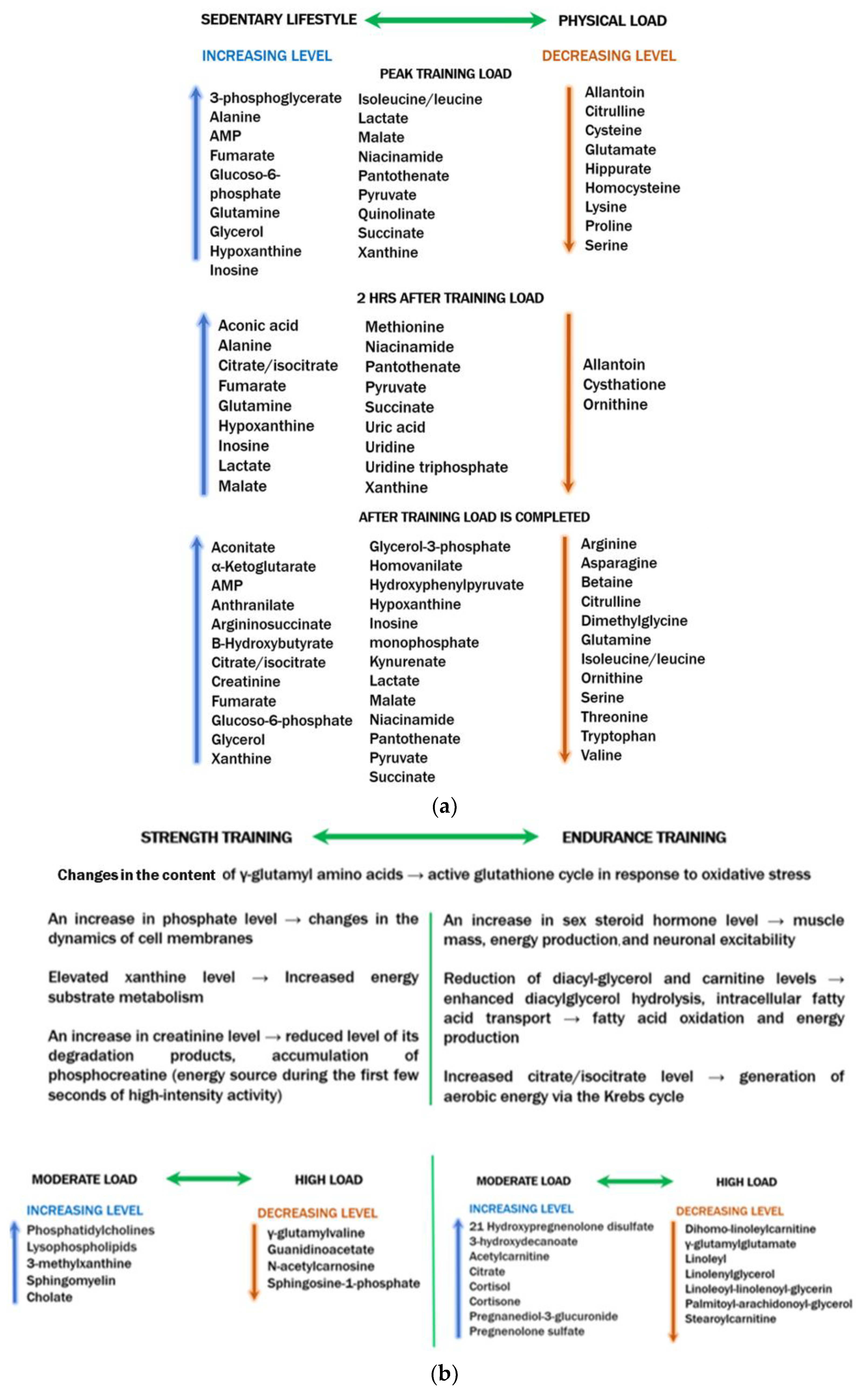
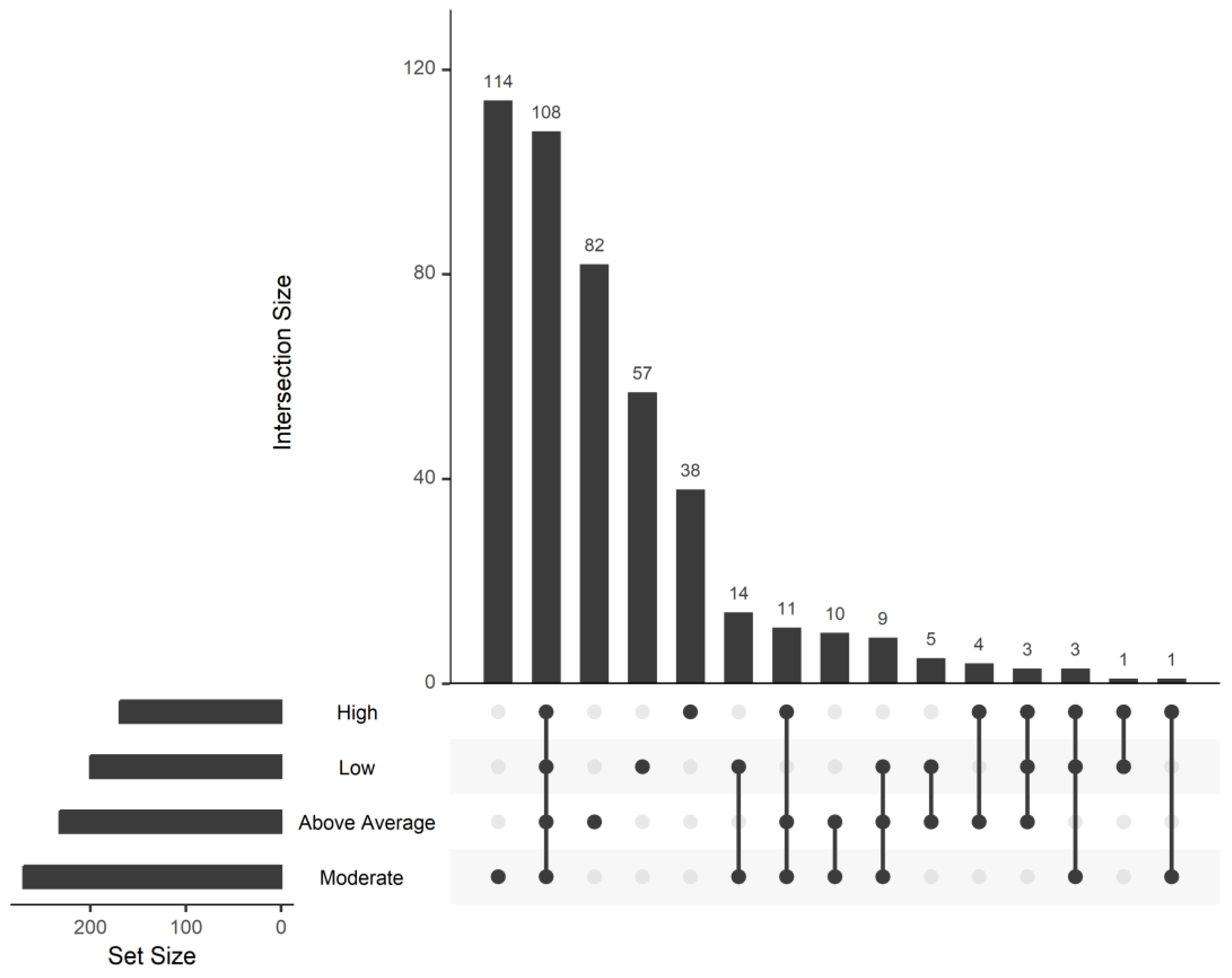
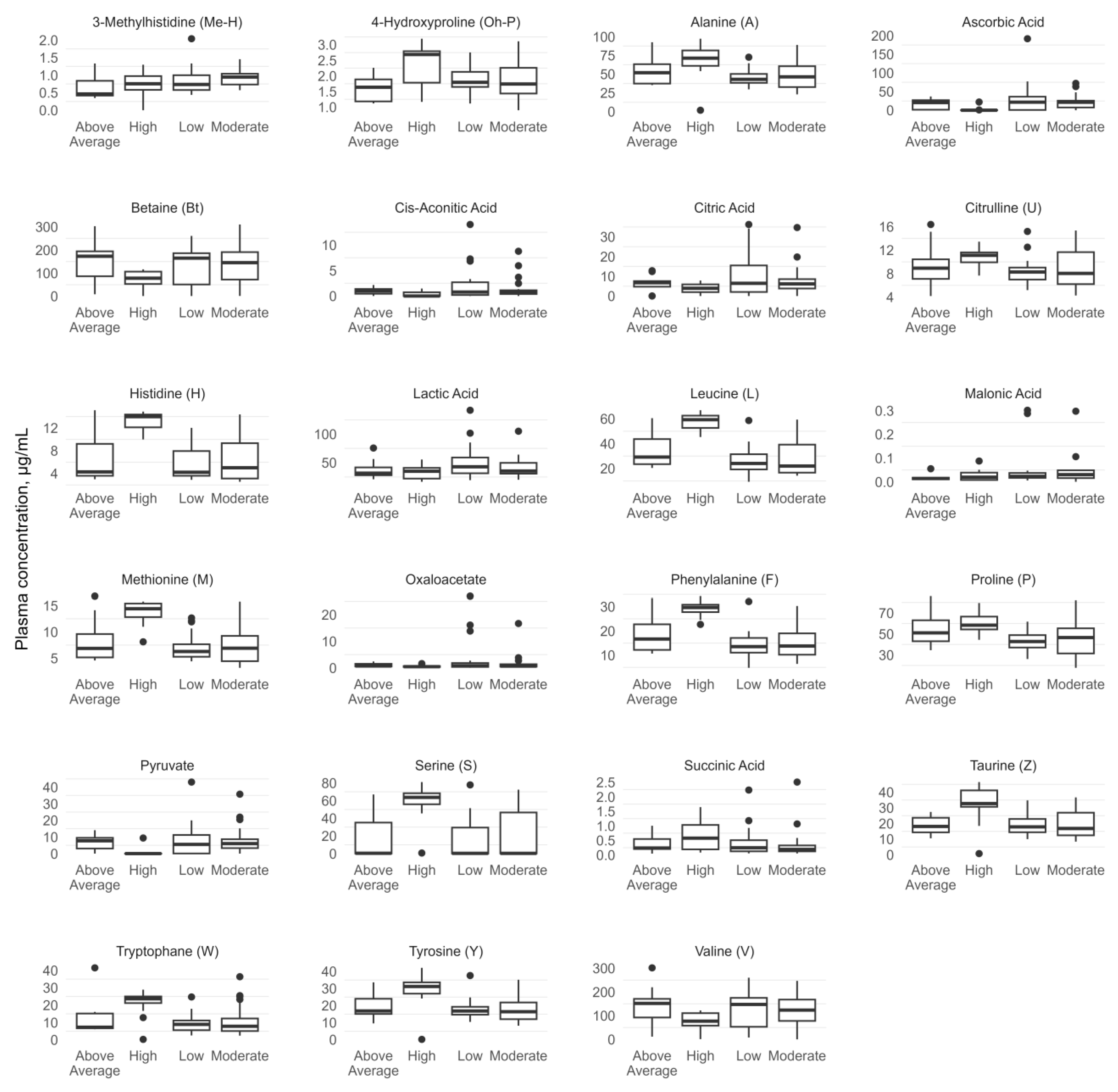
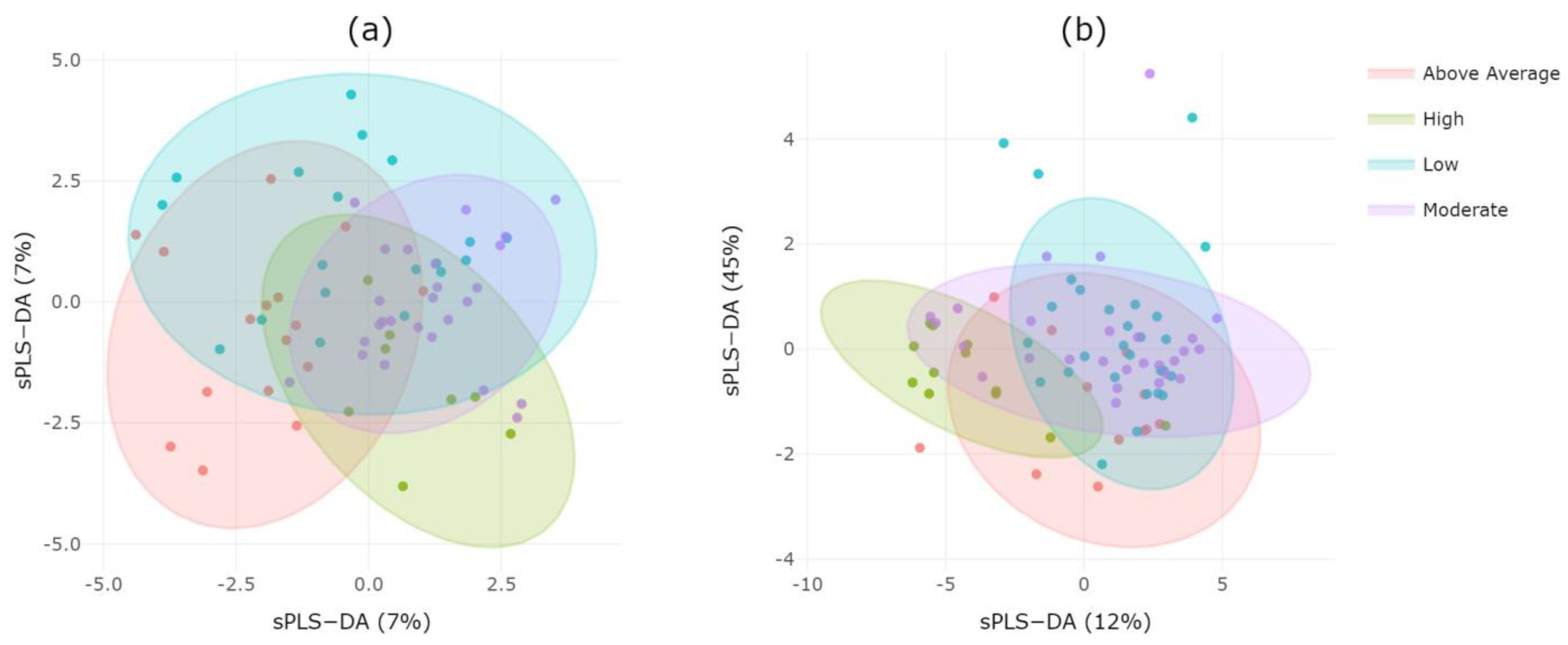
:1. Summary
2. Data Description
3. Methods
3.1. Ethics Statement
3.2. Subjects
- Sample information: the unique identifier of the study participant;
- Information about the anthropometric characteristics of the participant: the sex, age at the time of the examination, age at which their career began, and type of sport;
- Information about the clinical characteristics of the participant: allergies, infectious diseases, and sports injuries;
- Information about the training regime: main sports activities; the dynamics of sports results, number of training sessions per day during tapering and competition periods, number of rest days per week during tapering and competition periods, and training status self-assessment.
3.3. Preanalytical Stage of Analysis
3.4. HPLC-MS/MS Analysis
3.5. Data Analysis
4. User Comments
Supplementary Materials
Author Contributions
Funding
Institutional Review Board Statement
Informed Consent Statement
Data Availability Statement
Conflicts of Interest
References
- Malsagova, K.A.; Butkova, T.V.; Kopylov, A.T.; Izotov, A.A.; Rudnev, V.R.; Klyuchnikov, M.S.; Stepanov, A.A.; Kaysheva, A.L. Molecular Portrait of an Athlete. Diagnostics 2021, 11, 1095. [Google Scholar] [CrossRef] [PubMed]
- Bongiovanni, T.; Pintus, R.; Dessì, A.; Noto, A.; Sardo, S.; Finco, G.; Corsello, G.; Fanos, V. Sportomics: Metabolomics Applied to Sports. The New Revolution? Eur. Rev. Med. Pharmacol. Sci. 2019, 23, 11011–11019. [Google Scholar] [CrossRef]
- Nieman, D.C.; Gillitt, N.D.; Knab, A.M.; Shanely, R.A.; Pappan, K.L.; Jin, F.; Lila, M.A. Influence of a Polyphenol-Enriched Protein Powder on Exercise-Induced Inflammation and Oxidative Stress in Athletes: A Randomized Trial Using a Metabolomics Approach. PLoS ONE 2013, 8, e72215. [Google Scholar] [CrossRef]
- Pechlivanis, A.; Kostidis, S.; Saraslanidis, P.; Petridou, A.; Tsalis, G.; Mougios, V.; Gika, H.G.; Mikros, E.; Theodoridis, G.A. 1H NMR-Based Metabonomic Investigation of the Effect of Two Different Exercise Sessions on the Metabolic Fingerprint of Human Urine. J. Proteome Res. 2010, 9, 6405–6416. [Google Scholar] [CrossRef]
- Nieman, D.C.; Gillitt, N.D.; Henson, D.A.; Sha, W.; Shanely, R.A.; Knab, A.M.; Cialdella-Kam, L.; Jin, F. Bananas as an Energy Source during Exercise: A Metabolomics Approach. PLoS ONE 2012, 7, e37479. [Google Scholar] [CrossRef]
- Pintus, R.; Bongiovanni, T.; Corbu, S.; Francavilla, V.C.; DessÌ, A.; Noto, A.; Corsello, G.; Finco, G.; Fanos, V.; Marincola, F.C. Sportomics in Professional Soccer Players: Metabolomics Results during Preseason. J. Sports Med. Phys. Fit. 2021, 61, 324–330. [Google Scholar] [CrossRef] [PubMed]
- Schader, J.F.; Haid, M.; Cecil, A.; Schoenfeld, J.; Halle, M.; Pfeufer, A.; Prehn, C.; Adamski, J.; Nieman, D.C.; Scherr, J. Metabolite Shifts Induced by Marathon Race Competition Differ between Athletes Based on Level of Fitness and Performance: A Substudy of the Enzy-MagIC Study. Metabolites 2020, 10, 87. [Google Scholar] [CrossRef] [PubMed]
- Koay, Y.C.; Stanton, K.; Kienzle, V.; Li, M.; Yang, J.; Celermajer, D.S.; O’Sullivan, J.F. Effect of Chronic Exercise in Healthy Young Male Adults: A Metabolomic Analysis. Cardiovasc. Res. 2021, 117, 613–622. [Google Scholar] [CrossRef]
- Cao, B.; Liu, S.; Yang, L.; Chi, A. Changes of Differential Urinary Metabolites after High-Intensive Training in Teenage Football Players. BioMed Res. Int. 2020, 2020, 2073803. [Google Scholar] [CrossRef]
- Pechlivanis, A.; Papaioannou, K.G.; Tsalis, G.; Saraslanidis, P.; Mougios, V.; Theodoridis, G.A. Monitoring the Response of the Human Urinary Metabolome to Brief Maximal Exercise by a Combination of RP-UPLC-MS and 1H NMR Spectroscopy. J. Proteome Res. 2015, 14, 4610–4622. [Google Scholar] [CrossRef]
- Lewis, G.D.; Farrell, L.; Wood, M.J.; Martinovic, M.; Arany, Z.; Rowe, G.C.; Souza, A.; Cheng, S.; McCabe, E.L.; Yang, E.; et al. Metabolic Signatures of Exercise in Human Plasma. Sci. Transl. Med. 2010, 2, 33ra37. [Google Scholar] [CrossRef]
- Al-Khelaifi, F.; Diboun, I.; Donati, F.; Botrè, F.; Alsayrafi, M.; Georgakopoulos, C.; Suhre, K.; Yousri, N.A.; Elrayess, M.A. A Pilot Study Comparing the Metabolic Profiles of Elite-Level Athletes from Different Sporting Disciplines. Sports Med.-Open 2018, 4, 2. [Google Scholar] [CrossRef]
- Lu, W.; Bennett, B.D.; Rabinowitz, J.D. Analytical Strategies for LC–MS-Based Targeted Metabolomics. J. Chromatogr. B 2008, 871, 236–242. [Google Scholar] [CrossRef]
- Alghannam, A.F.; Ghaith, M.M.; Alhussain, M.H. Regulation of Energy Substrate Metabolism in Endurance Exercise. Int. J. Environ. Res. Public. Health 2021, 18, 4963. [Google Scholar] [CrossRef]
- Pettersson, J.; Hindorf, U.; Persson, P.; Bengtsson, T.; Malmqvist, U.; Werkström, V.; Ekelund, M. Muscular Exercise Can Cause Highly Pathological Liver Function Tests in Healthy Men. Br. J. Clin. Pharmacol. 2008, 65, 253–259. [Google Scholar] [CrossRef] [PubMed]
- Clarkson, P.M.; Kearns, A.K.; Rouzier, P.; Rubin, R.; Thompson, P.D. Serum Creatine Kinase Levels and Renal Function Measures in Exertional Muscle Damage. Med. Sci. Sports Exerc. 2006, 38, 623–627. [Google Scholar] [CrossRef] [PubMed]
- Luoto, R.; Ruuskanen, O.; Ihalainen, J.K.; Pekkala, S.; Hintikka, J.; Kanerva, N.; Waris, M.; Heinonen, O.J.; Valtonen, M. Inflammatory Biomarkers in Elite Cross-Country Skiers after a Competition Season: A Case–Control Study. J. Sci. Sport Exerc. 2023, 5, 254–262. [Google Scholar] [CrossRef]
- Muscella, A.; My, G.; Okba, S.; Zangla, D.; Bianco, A.; Marsigliante, S. Effects of Training on Plasmatic Cortisol and Testosterone in Football Female Referees. Physiol. Rep. 2022, 10, e15291. [Google Scholar] [CrossRef]
- Gonzalez-Bono, E.; Salvador, A.; Serrano, M.A.; Ricarte, J. Testosterone, Cortisol, and Mood in a Sports Team Competition. Horm. Behav. 1999, 35, 55–62. [Google Scholar] [CrossRef]
- Forte, P.; Branquinho, L.; Ferraz, R. The Relationships between Physical Activity, Exercise, and Sport on the Immune System. Int. J. Environ. Res. Public. Health 2022, 19, 6777. [Google Scholar] [CrossRef]
- Gleeson, M. Immune function in sport and exercise. J. Appl. Physiol. 2007, 103, 693–699. [Google Scholar] [CrossRef] [PubMed]
- Hadžović–Džuvo, A.; Valjevac, A.; Lepara, O.; Pjanić, S.; Hadžimuratović, A.; Mekić, A. Oxidative Stress Status in Elite Athletes Engaged in Different Sport Disciplines. Bosn. J. Basic Med. Sci. 2014, 14, 56–62. [Google Scholar] [CrossRef] [PubMed]
- Marin, D.P.; Bolin, A.P.; Campoio, T.R.; Guerra, B.A.; Otton, R. Oxidative Stress and Antioxidant Status Response of Handball Athletes: Implications for Sport Training Monitoring. Int. Immunopharmacol. 2013, 17, 462–470. [Google Scholar] [CrossRef]
- Havermale, L.A. Nutrition Knowledge of Collegiate Athletes in Endurance and Non-Endurance Sports; Southern Illinois University: Carbondale, IL, USA, 2017; 38p. [Google Scholar]
- Stepanov, A.A.; Malsagova, K.A.; Kopylov, A.T.; Rudnev, V.R.; Karateev, D.E.; Markelova, E.I.; Luchikhina, E.L.; Borisova, E.E.; Kaysheva, A.L. Determination of Heterogeneous Proteomic and Metabolomic Response in Anti-TNF and Anti-IL-6 Treatment of Patients with Rheumatoid Arthritis. Life 2023, 13, 596. [Google Scholar] [CrossRef] [PubMed]
- Bittremieux, W.; Walzer, M.; Tenzer, S.; Zhu, W.; Salek, R.M.; Eisenacher, M.; Tabb, D.L. The Human Proteome Organization–Proteomics Standards Initiative Quality Control Working Group: Making Quality Control More Accessible for Biological Mass Spectrometry. Anal. Chem. 2017, 89, 4474–4479. [Google Scholar] [CrossRef]
- Petrovskiy, D. Proteomic and Metabolomic Analyses of Blood Samples of Highly Trained Athletes. Figshare 2023. [Google Scholar] [CrossRef]
- Deutsch, E.; Lane, L.; Overall, C.; Bandeira, N.; Baker, M.; Pineau, C.; Moritz, R.; Corrales, F.; Orchard, S.; Eyk, J.; et al. Human Proteome Project Mass Spectrometry Data Interpretation Guidelines 3.0. J. Proteome Res. 2019, 2019, 9b00542. [Google Scholar] [CrossRef]
- Kopylov, A.T.; Petrovsky, D.V.; Stepanov, A.A.; Rudnev, V.R.; Malsagova, K.A.; Butkova, T.V.; Zakharova, N.V.; Kostyuk, G.P.; Kulikova, L.I.; Enikeev, D.V.; et al. Convolutional Neural Network in Proteomics and Metabolomics for Determination of Comorbidity between Cancer and Schizophrenia. J. Biomed. Inform. 2021, 122, 103890. [Google Scholar] [CrossRef]
- Petrovsky, D.V.; Pustovoyt, V.I.; Nikolsky, K.S.; Malsagova, K.A.; Kopylov, A.T.; Stepanov, A.A.; Rudnev, V.R.; Balakin, E.I.; Kaysheva, A.L. Tracking Health, Performance and Recovery in Athletes Using Machine Learning. Sports 2022, 10, 160. [Google Scholar] [CrossRef]
- Petrovsky, D.V.; Kopylov, A.T.; Rudnev, V.R.; Stepanov, A.A.; Kulikova, L.I.; Malsagova, K.A.; Kaysheva, A.L. Managing of Unassigned Mass Spectrometric Data by Neural Network for Cancer Phenotypes Classification. J. Pers. Med. 2021, 11, 1288. [Google Scholar] [CrossRef]
- Ahsan, M.M.; Luna, S.A.; Siddique, Z. Machine-Learning-Based Disease Diagnosis: A Comprehensive Review. Healthcare 2022, 10, 541. [Google Scholar] [CrossRef] [PubMed]
- Choi, S.B.; Kim, W.J.; Yoo, T.K.; Park, J.S.; Chung, J.W.; Lee, Y.; Kang, E.S.; Kim, D.W. Screening for Prediabetes Using Machine Learning Models. Comput. Math. Methods Med. 2014, 2014, 618976. [Google Scholar] [CrossRef] [PubMed]
- Hsieh, C.-H.; Lu, R.-H.; Lee, N.-H.; Chiu, W.-T.; Hsu, M.-H.; Li, Y.-C.J. Novel Solutions for an Old Disease: Diagnosis of Acute Appendicitis with Random Forest, Support Vector Machines, and Artificial Neural Networks. Surgery 2011, 149, 87–93. [Google Scholar] [CrossRef]
- Balasubramanian, J.B.; Boes, R.D.; Gopalakrishnan, V. A Novel Approach to Modeling Multifactorial Diseases Using Ensemble Bayesian Rule Classifiers. J. Biomed. Inform. 2020, 107, 103455. [Google Scholar] [CrossRef]


| Kind of Sport | Load Type | Intensity | Number of Athletes |
|---|---|---|---|
| Sailing | Strength Endurance | High | 11 |
| Kayaking and Canoeing | 1 | ||
| Freestyle Wrestling | Endurance | Above Average | 20 |
| Sambo | Speed-strength | Moderate | 2 |
| Figure Skating | Strength Endurance | 1 | |
| Rowing | Technical | 16 | |
| Beach Soccer | 4 | ||
| Football | 3 | ||
| Ski Race | 6 | ||
| Biathlon | Endurance | Low | 7 |
| Greco-Roman Wrestling | Technical | 20 | |
| Athletics | 2 |
| Intensity | Number of Proteins | Number of Proteins in the Group | |||
|---|---|---|---|---|---|
| Min | Max | Mean | SE | ||
| Above Average | 61 | 122 | 84.4 | 4.2 | 232 |
| High | 71 | 106 | 88.1 | 5.1 | 169 |
| Low | 61 | 103 | 81.5 | 2.3 | 200 |
| Moderate | 60 | 111 | 83.5 | 2.9 | 270 |
| Intensity | Number of Athletes | %, Men | Age, Years | Allergy, % | Bad Habits, % |
|---|---|---|---|---|---|
| High | 12 | 83 | 33.2 ± 6.3 | 33 | – |
| Moderate | 32 | 72 | 25.5 ± 2.8 | 13 | – |
| Above Average | 20 | 100 | 29.2 ± 2.5 | – | – |
| Low | 29 | 76 | 30.0 ± 3.7 | 7 | – |
Disclaimer/Publisher’s Note: The statements, opinions and data contained in all publications are solely those of the individual author(s) and contributor(s) and not of MDPI and/or the editor(s). MDPI and/or the editor(s) disclaim responsibility for any injury to people or property resulting from any ideas, methods, instructions or products referred to in the content. |
© 2024 by the authors. Licensee MDPI, Basel, Switzerland. This article is an open access article distributed under the terms and conditions of the Creative Commons Attribution (CC BY) license (https://creativecommons.org/licenses/by/4.0/).
Share and Cite
Malsagova, K.A.; Kopylov, A.T.; Pustovoyt, V.I.; Balakin, E.I.; Yurku, K.A.; Stepanov, A.A.; Kulikova, L.I.; Rudnev, V.R.; Kaysheva, A.L. Proteomic and Metabolomic Analyses of the Blood Samples of Highly Trained Athletes. Data 2024, 9, 15. https://doi.org/10.3390/data9010015
Malsagova KA, Kopylov AT, Pustovoyt VI, Balakin EI, Yurku KA, Stepanov AA, Kulikova LI, Rudnev VR, Kaysheva AL. Proteomic and Metabolomic Analyses of the Blood Samples of Highly Trained Athletes. Data. 2024; 9(1):15. https://doi.org/10.3390/data9010015
Chicago/Turabian StyleMalsagova, Kristina A., Arthur T. Kopylov, Vasiliy I. Pustovoyt, Evgenii I. Balakin, Ksenia A. Yurku, Alexander A. Stepanov, Liudmila I. Kulikova, Vladimir R. Rudnev, and Anna L. Kaysheva. 2024. "Proteomic and Metabolomic Analyses of the Blood Samples of Highly Trained Athletes" Data 9, no. 1: 15. https://doi.org/10.3390/data9010015