Article

Effects of Extreme-Ambient Temperatures in Silver Barb (Barbonymus gonionotus): Metabolic, Hemato-Biochemical Responses, Enzymatic Activity and Gill Histomorphology

1
Department of Genetics and Fish Breeding, Bangabandhu Sheikh Mujibur Rahman Agricultural University, Gazipur 1706, Bangladesh
2
Division of Stress Physiology and Molecular Biology, Institute of Climate Change and Environment, Bangabandhu Sheikh Mujibur Rahman Agricultural University, Gazipur 1706, Bangladesh
3
Department of Earth Sciences and Environment, Faculty of Science and Technology, Universiti Kebangsaan Malaysia (UKM), Bangi 43600, Selangor, Malaysia
4
Department of Fish Health Management, Sylhet Agricultural University, Sylhet 3100, Bangladesh
5
Department of Oceanography, Khulna Agricultural University, Khulna 9100, Bangladesh
*
Author to whom correspondence should be addressed.
Water 2024, 16(2), 292; https://doi.org/10.3390/w16020292
Submission received: 10 November 2023 / Revised: 21 December 2023 / Accepted: 26 December 2023 / Published: 15 January 2024
(This article belongs to the Special Issue Impact of Climate Changes on Marine Ecology and Fishes)

Abstract

:
A global interest in Silver Barb (Barbonymus gonionotus) cultivation has arisen because of a combination of declining wild stock and a steady increase in demand and market value. The purpose of the current investigation was to evaluate the impact of extreme water temperature changes on growth, hemato-biochemical responses, pepsin enzyme activity and gill histomorphology of B. gonionotus. Four distinct temperatures (22, 26, 30 and 34 °C) were applied in the laboratory (22, 26, 30 and 34 °C) in triplicate glass aquariums (150 L each). At 30 and 22 °C, respectively, the highest and the lowest length and weight gain, specific, daily and relative growth rates were noted. At 30 °C the lowest FCR was recorded (1.42 ± 0.04). The values related to hematology and biochemistry were found to be within normal ranges; nevertheless, there was a notable variation in the parameters when the temperature changed. Hematological results revealed that RBC, HB, PLT, HCT and PMV levels were significantly higher in groups 30 °C with respect to others. It was evident that cold and heat shock stress was present due to the decline in hematocrit levels and rise in WBC values. Regarding the biochemical responses, the level of triglycerides, cholesterol, AP, Ca+, HDL, and HDL/LDL ratio increased significantly higher at 30 °C. Glucose, total protein, albumin, Na+, K+, Cl−1, AST, bilirubin, uric acid, and LDL levels were significantly higher at 22 °C compared to other temperatures. There was a higher pepsin activity between 26 and 30 °C while the digestive somatic index (IDS) was disproportionate. Histological examination demonstrated the well-shaped gill tissues at 26 and 30 °C compared to distinctive pathology in other temperature treatments. As an end note, the results of the experiment indicated that B. gonionotus should be cultured at 26–30 °C to maximize the overall productivity and the health condition of this economically important fish species.

1. Introduction

The threat posed by global climate change to natural systems and their species is grave and is only becoming worse. More specifically, the changes in temperature patterns have a severe impact on aquatic habitats [1,2]. Stakeholders and researchers in fisheries and aquaculture are also somewhat concerned about it. It is anticipated that temperature trends will occur more intensively and frequently than in the past due to the significant changes in recent decades at both the global and regional levels [3,4]. In tropical and subtropical areas, temperature variations brought on by climate change are predicted to have an impact on teleosts’ physiological processes [5]. Fish growth, physiology [6,7], metabolism [8], and immune responses [9] are all hampered by temperature changes in the water, which causes fish abundance to decline in extreme cases and cause distribution shifts [10,11]. Because aquaculture fish cannot change their distribution, the effects on them are little understood, yet they are crucial to maintaining aquaculture’s sustained contribution to food security worldwide [12].
One of the most important aspects in assessing the economic feasibility of commercial fish culture is growth, which is known to be influenced by a number of biotic (such as food, predation, and population dynamics) and abiotic (physical and chemical characteristics of the water) variables [6,13,14,15]. Because ectothermic animals are extremely sensitive to temperature fluctuations, low temperatures cause their metabolic rates to be inhibited, which lowers aquaculture productivity [16]. Alternatively, higher temperatures are said to speed up metabolism, although fish may experience physiological stress if the temperature rises too high [17]. Fish digestive enzymes have received less research attention, despite the fact that bony fishes have the same array of digestive enzymes as other vertebrates. Pepsins are vital digestive enzymes that play a major role in the breakdown of proteins in fish diets. Pepsin enzyme levels in fish can be affected by a variety of native and exogenous variables, including the fish’s age, the kind of food it consumes, the time of year, and the temperature at which it acclimatizes. However, the fact that enzyme activity is greatly dependent on temperature, as well as on the amount and makeup of food, presents one of the biggest challenges to comprehending the exerted action (the cumulative conversion catalyzed by enzymes) [18].
The hematological and biochemical components are significantly impacted by the temperature of the living medium, and fluctuations within the ideal range restrict the fish’s physiological well-being [19]. Hemato-biochemical indices are often employed to assess fish health [20] and can offer details on the physiological reactions to environmental modifications that impact homeostasis [6,21]. Variations in the chemical properties of fish blood could be signs of temperature stress and serve as possible indicators of the physiological well-being of aquatic animals [22]. Fish essential organs like gills undergo cellular and histological changes and may serve as an effective biomarker for various exogenous and endogenous stressors connected to their drinking water [23,24]. Bangladesh is regarded as one of the tropical nations most vulnerable to global warming, and the country’s freshwater fishing resources and aquaculture industries are severely impacted by the phenomenon.
Among the several species, one that stands out is the Silver barb, Barbonymus gonionotus, for its economic significance, commercial appeal, and widespread use as a food source [25]. Its great taste and high productivity have made it popular [26]. In addition, it adapts favorably to relatively low-cost and easy management techniques [27] and thus establishing it as a most suitable species for fish farmers. Recently, the practice of intensive culture of this species has been gaining popularity in fish-producing countries like Bangladesh [28]. However, questions about the species’ metabolic features and capacity to handle stressors from the surroundings have gotten little consideration. To date, research on Silver barb has concentrated on pesticide tolerance [29,30], dietary replacement [31,32], growth, nutrition [28,32] and heavy metal tolerance [33,34]. There is a lack of information on the response of physiological, hematological, biochemical responses and gill histomorphology of Silver barb encountering extreme temperature events has not been comprehensively studied. Therefore, the current experiment aimed to identify the impact of extreme water temperature changes on growth performances, hemato-biochemical responses, pepsin enzyme activity and the gill histomorphology of B. gonionotus.

2. Methodology

2.1. Experimental Fish Collection, Transportation and Conditioning

The experiment was carried out at the laboratory and hatchery complex of the Department of Genetics and Fish Breeding, Bangabandhu Sheikh Mujibur Rahman Agricultural University (BSMRAU), Gazipur. the specimens of B. gonionotus (11.27 ± 0.12 cm TL and 12.53 ± 0.82 g BW) were collected from local hatcheries of Mymensingh, Bangladesh and transported to the Genetics and Fish Breeding laboratory of BSMRAU, Bangladesh.
The fish were kept in a 4000 L cemented stocking tank at 26 ± 0.5 °C for 21 days before starting experiments to recover from the impacts of handling and transportation stress and adapt to the new environment and food. Fish were given enough commercial starter feed twice daily. Every attempt was made to ensure the well-being of the animals, minimizing any potential distress.

2.2. Experimental Design

Twelve aquaria (80 cm × 45 cm × 40 cm) each were filled with 150 L of freshwater in the laboratory of the Department of Genetics and Fish Breeding, BSMRAU, Bangladesh. After setting, fifteen individuals of B. gonionotus were placed within every aquarium and enough aeration was provided. The fish were exposed to four temperature treatments: 22, 26, 30, and 34 °C for 60 days. We considered 26 °C as the controlled temperature. Each temperature was kept constant in a series of three replicate tanks (3 tanks × 4 temperatures) (Figure 1). Temperature adjustments were initiated for each of the experimental treatments at 2 °C day−1 by using a water heater (E-JET Heater 200W, Penang, country of origin: Malaysia) and a cooler (HS-28A 250–1200 L/H, Guangdong Hailea Group Co., Ltd., Chaozhou, country of origin: China) until the investigational temperature reached to the lowest possible at 22 °C and the highest at 34 °C. A 12:12-h light:dark photoperiod was maintained. Throughout the trial, 20% of the water was replaced daily. Fish were sampled and data were recorded every 15 days. A commercial pellet diet (moisture: 12.98%, protein: 31.20%, lipid: 6.72%, ash: 13.22%, fiber: 11.58%, and NFE: 24.30%) was supplied to the fish twice a day until they were satiated at 09.00 and 16.00 h. Besides, feces of experimental fish and uneaten feedstuff were removed daily.

2.3. Measurements of Water Quality

The water temperature and pH were tested daily while total hardness and total ammonia nitrogen (NH3-N) were determined weekly. Every measurement was made at 9:00 a.m. A YSI 59 Multiparameter Water Quality Probe (Yellow Springs Instrument Company, Yellow Springs, OH, country of origin: USA) was used to measure both the temperature and pH of water. The salicylate method (HachTM method 8155) was used to measure NH3-N, and the titration (La Motte Chemical test kit, model WAT-DR) method was used to assess total hardness.

2.4. Evaluation of the Parameters of Growth Performance

After 60 days of experiment, all fish were tallied, measured, and weighed. The average weight of each group was determined by dividing the total weight by the number of fish. Various growth parameters were computed by using the formulas:
Body weight gain (BWG, g) = (W2W1) × n
Food consumption (FC, gday−1) = (g food consumed × day−1)
Food conversion ratio (FCR) = F/W2W1
Food conversion efficiency (FCE) = (gain in fish mass × g food consumed−1)
Specific growth rate (SGR, %day−1) = 100 × (lnW2 − lnW1/t
Relative growth rate (RGR, %) = 100 × (W2W1)/W1
Daily growth rate (DGR, %) = 100 × (W2W1)/t
Survival rate % = [Final number of fish/initial number of fish] × 100
Here, F is the entire amount of food consumed during the trial period; W1 and W2 are the initial (0 d) and final (60 d) mean individual weights for each treatment (to the nearest 0.01 g), n is the final no. of fish, and t is the duration of the investigational period.

2.5. Hemato-Biochemical Responses

After the completion of the experimental period, the fish were kept fasting for 48 h, then removed from the holding tank and dipped in anesthetic solution [α-methyl quinoline (Transmore®; Nika Trading, Puchong, country of origin: Malaysia) (0.22 mL L−1) until ventilatory movements stopped. After that, the fish were placed on a surgical table. Five fish had blood samples taken from their caudal veins (non-lethal sampling) using a disinfected 2.5 mL plastic syringe and shifted into 2 tubes: the first aliquot was collected in EDTA containing tube (1.26 mg/0.6 mL) and kept for hematological evaluations at 4 °C, while the other portion was leftover for 30 min to clot, centrifuged at 12,000× g at 4 °C for 15 min, and the collected serum was kept for further analysis at −20 °C. An upgraded Neubaur hemocytometer was used to count the total red blood cells (RBCs) [35]. Cyanmethemoglobin and microhematocrit techniques were used to determine hemoglobin (Hb) and hemocrit (Hct) [36]. With some modifications, a Neubaur hemocytometer was used to count the total white blood cells (WBCs) [37].
For biochemical analysis, a clear fluid: the serum was pipetted into a 1.5 mL sterile Eppendorf tube that had been cleaned and sterilized [38]. For each sample, 800 μL of serum was applied to numbered sample wells. Albumin (ALB), total protein (TP), globulin (GLB), alkaline/globulin ratio (A/G ratio), alkaline phosphatase (AP), alanine transaminase (ALT), and aspartate aminotransferase (AST) were measured using assay kits (Biosino Bio-Technology and Science Inc., Beijing, China) [39].

2.6. Pepsin Enzymatic Activity

After collecting blood samples, the total gut contents were collected from each group. To account for the potential impact of variations in individual body mass, the digestive somatic index (IDS) was determined. Fish were weighed (M) and had their entire gut taken (from the esophagus to the anus) by making an incision at the esophagus and cloaca to calculate the IDS [40]. Then it was cut longitudinally and thoroughly rinsed in ice-cold 0.1 mol L−1 phosphate-buffered saline (PBS, pH 7.4). The intestine was blotted dry with filter paper after rinsing, weighed (MG) and stored at −20 °C until analyzed. Every attempt was made to reduce suffering.
The IDS (g) was calculated as: IDS = 100 MG M−1
After carefully unfreezing, the guts were separated and weighed using a needle. Individual guts were homogenized by glass and electric Teflon homogenizer (Polytron, Heidolph RZR 1, country of origin: Germany) in 20 volumes (v/w) of ice-cold 50 mM Tris-HCl buffer solution with pH 7.4 following standard procedure [13]. The Bradford assay was used to assess the total protein content of the intestinal supernatant using the BioRadR assay kit [41]. Pepsin’s specific activity was measured in relation to the extracts’ soluble protein content. Utilizing 2% hemoglobin in 0.06 N HCl as a substrate, the activity was measured, and the result was derived from the work of Natali et al. [42]. The test is based on Anson’s stop-point assay for hemoglobin degradation [43]. Tris-HCl (pH 7.4) was the buffer that was employed. The mixture contained 0.5 mL of hemoglobin, 1 mL of TCA, and 0.1 mL of crude enzyme in a 2% (w/v) solution. There are two steps in the process: one for blank tubes and another for unknown samples. Both the samples and the blanks underwent double analysis. First, added 500 µL of the substrate (2% bovine hemoglobin) and 100 µL of crude enzyme extract to each tube. Additionally, 500 µL of the substrate (2% bovine hemoglobin) was obtained for blank tubes. After all solutions were incubated for 10 min at 37 °C in a hot air oven, the reaction was halted by adding 1 milliliter of 5% trichloroacetic acid (TCA) and 100 microliters of crude enzyme extract to a blank tube. For five minutes, all the samples and blanks were kept at room temperature. Following a 5-min centrifugation at 12,000× g, the samples were measured using an Ultra Spec 2000 Pharmacia Biotech UV/VS spectrophotometer, and absorbance was measured at 280 nm. Units per milligram of protein (U mg protein−1) were used to express the specific enzyme activity as:
[Absorbance value at 280 (supernatant) − Absorbance value at 280 (blank)] × 1000/(10 min × mg protein)

2.7. Histological Examination

Following the 60-day experimental period, the spinal cords of two fish from each tank were severed immediately behind the opercula. Once the fish was deceased, the gills were preserved in 10% neutral buffered formalin. The gill tissues were then embedded in paraffin wax, and sections measuring 3–5 μm thick were cut. These sections were subsequently stained with hematoxylin and eosin (H&E) and examined using a light microscope. The purpose of this examination was to identify any histological abnormalities or lesions present in the gills [44].

2.8. Statistical Analysis

The data (mean ± SE) were analyzed by applying a one-way analysis of variance (ANOVA) followed by Duncan’s multiple range test to compare the means between experimental treatments. Before conducting any analysis, we checked the data for normality using the Shapiro–Wilk test and assessed homogeneity of variance using Levene’s test. Minitab version 18 and Origin software version 8 were used for statistical analyses. p < 0.05 was the accepted significance level.

3. Results

3.1. Measurements of Water Quality Parameters

The average values with standard error (SE) of water quality parameters measured in the experimental tanks in the study period are shown in Table 1. These parameters remained steady at the designated temperature levels, and there were no noteworthy differences observed (p > 0.05) in any of the parameters. The recorded values fall within the appropriate ranges for cultivating this species [45].

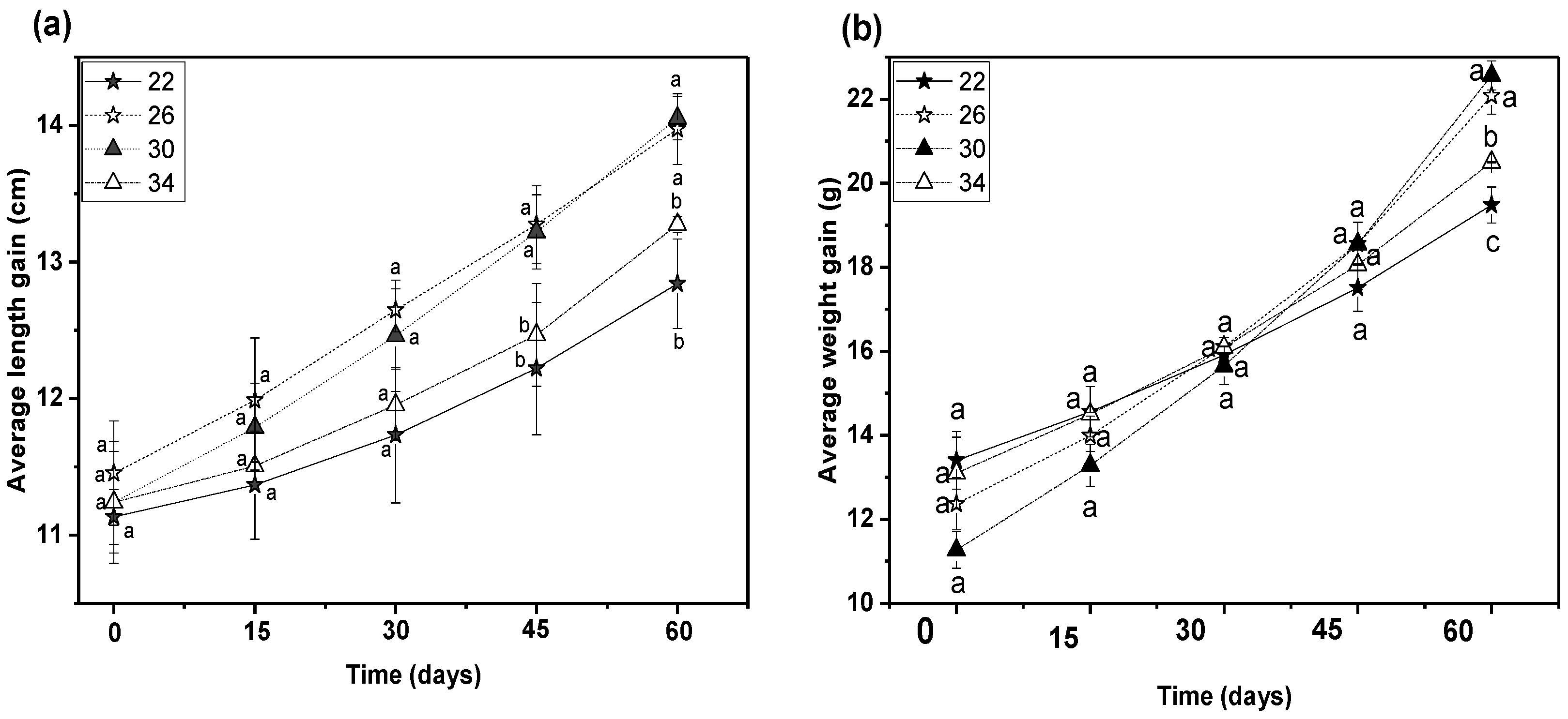
3.2. Growth Performances Study

In general, the four temperatures showed similar mean beginning length and weight values (Figure 2); the mean total length was 11.27 ± 0.12 cm, and individual weight was approximately 12.53 ± 0.82 g. In every treatment, there was 100% fish survival. The graphs show the variation of exposed temperatures determines growth, where the observed average total length and body weight of Silver barb up to 60 days of trial with a commercial diet. After 60 days, Growth at 30 °C and 26 °C was significantly higher than at other temperature conditions (p < 0.05, Figure 2). There was no significant difference in mean body weights between fish retained at 26 and 30 °C; however, the weight observed in both treatments was significantly higher than fish held at 22 and 34 °C (p < 0.05, Figure 2).
This 60-day experiment discovered that the water temperature had significant effects on the BWG, FCE, FCR, DGR, SGR, and RGR of the fish. The highest values of FTL, FBW, and BWG have been observed in fish reared at 30 °C (14.05 ± 1.66, 22.57 ± 4.35, and 11.30 ± 1.96, respectively). FCR values were significantly higher in fish reared at 22 and 34 °C (1.83 ± 0.05 and 1.75 ± 0.02) compared with those at 26 and 30 °C (1.51 ± 0.03 and 1.42 ± 0.04; p < 0.05, Table 2). When the temperature was raised from 22 °C to 30 °C, the values steadily fell, but substantially increased when the temperature was elevated to 34 °C. Overall, the group raised at 30 °C demonstrated the best FCR performance. On the other hand, the ideal temperature for FCE was 30 °C and estimated to be 0.70 ± 0.01.
Temperature has a strong influence on SGR. When the water temperature was altered from 22 °C to 30 °C, the SGR increased significantly (p < 0.05) and then reduced when the temperature was elevated from 30 °C to 34 °C (Table 2). The SGR value at 30 °C (1.17 ± 0.06) was significantly higher (p < 0.05) than in other treatments and the lowest SGR value was obtained in the treatment exposed at 22 °C (0.63 ± 0.07). Similarly, RGR and DGR also differed with temperature variations and significant variations were observed among the treatments used (p < 0.05, Table 2). The highest RGR and DGR were found in the 30 °C treated group (101.32 ± 6.99 and 113.00 ± 19.59, respectively), whereas the lowest RGR was at 34 °C (45.99 ± 5.81) and DGR (40.53 ± 9.49) was found at 22 °C.

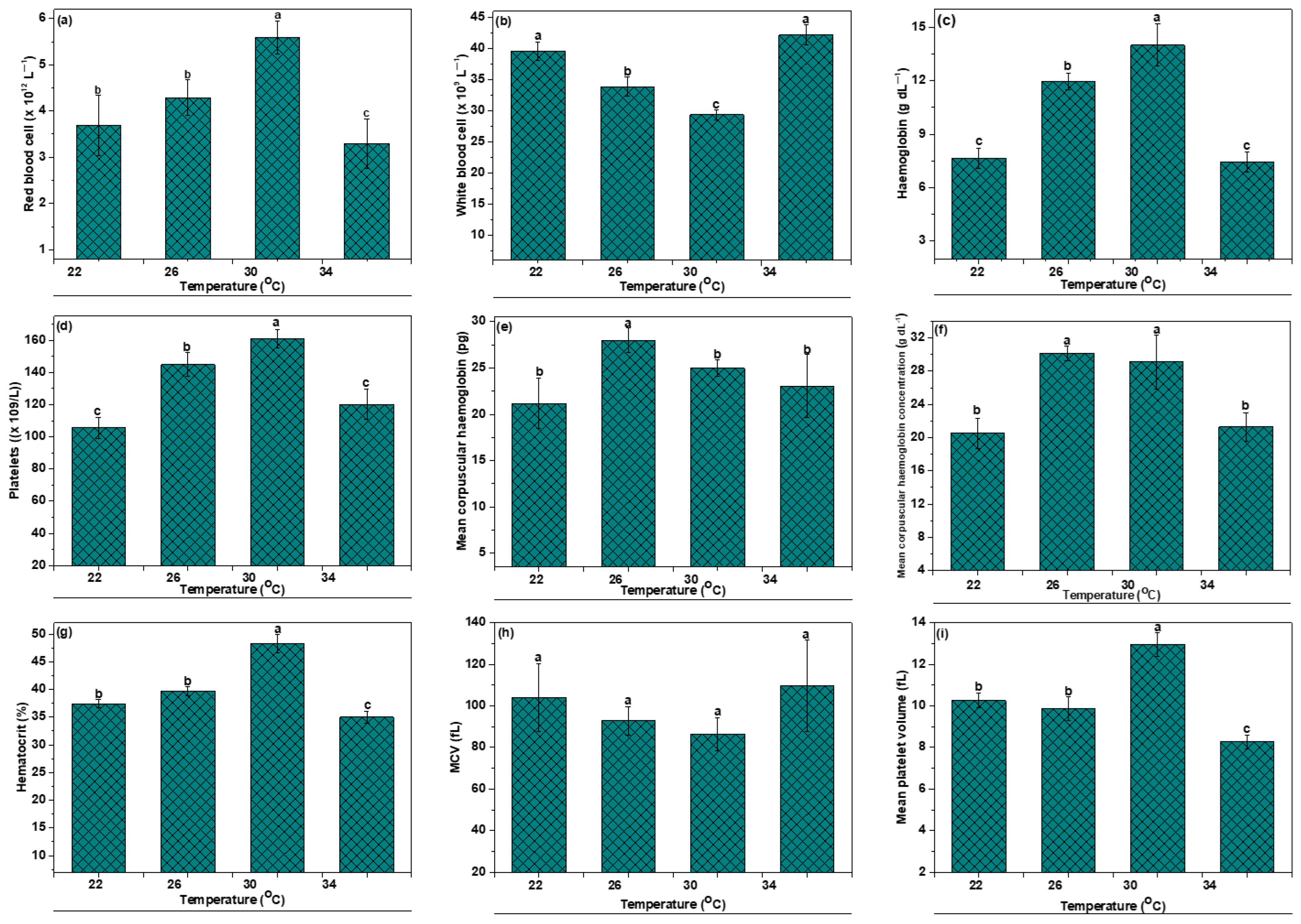
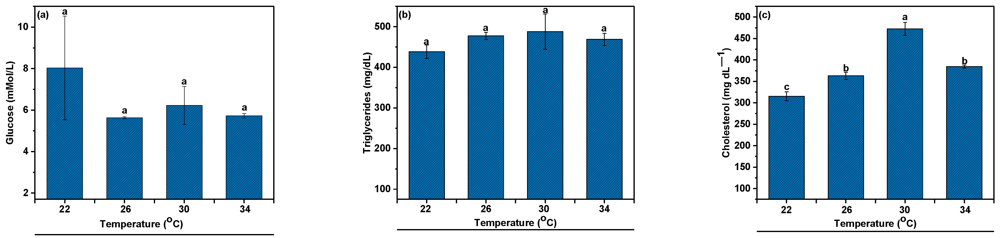
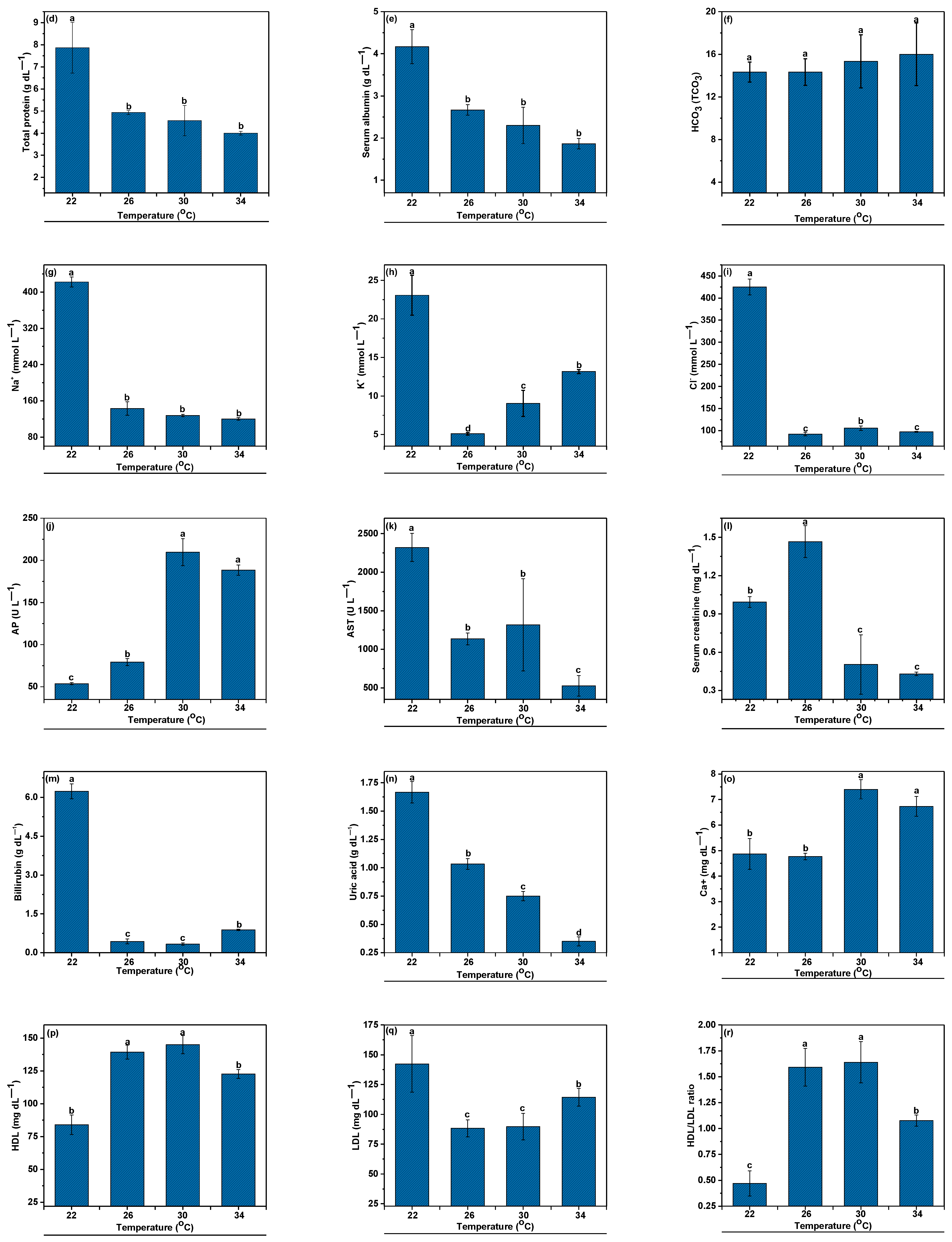
3.3. Hematological Indices

The one-way ANOVA analysis revealed that the experimental temperatures had a notable impact on different hematological parameters (Figure 3, p < 0.05). RBC, HGB, PLT, HCT and PMV levels were significantly higher (p < 0.05) in groups 30 °C with respect to other groups. Nonetheless, the level of WBCs (×109/L) followed the opposite trend with the RBCs and HGB throughout the experiment clearly indicating cold and heat shock stress. The MCH value dropped in hypochromic anemia due to a decrease in cell hemoglobin. Nonetheless, the MCH rose in the macrocytic state.
Figure 4 displays the computed biochemical parameters regarding temperature impacts. In the present study, the glucose, triglycerides and HCO3 levels were not affected by temperature change significantly (p > 0.05). Cholesterol, AP, Calcium and HDL levels showed higher values at 30 °C with respect to other temperature groups (p < 0.05). However, values of total serum proteins, albumin, Na+, K+, Cl, AST, bilirubin, uric acid, and LDL increased significantly from the threshold level indicating that the fish suffered from stress. Changes in the differences in total protein show changes in the relative protein fractions. Albumin showed a similar trend, increasing by more than 100% from the lowest at 34 °C representing the main contributor to rising levels of total proteins. The one-way ANOVA analysis proved that the change in the rearing temperature had a significant effect on all the biochemical parameters except glucose, triglycerides and HCO3 (p < 0.05).

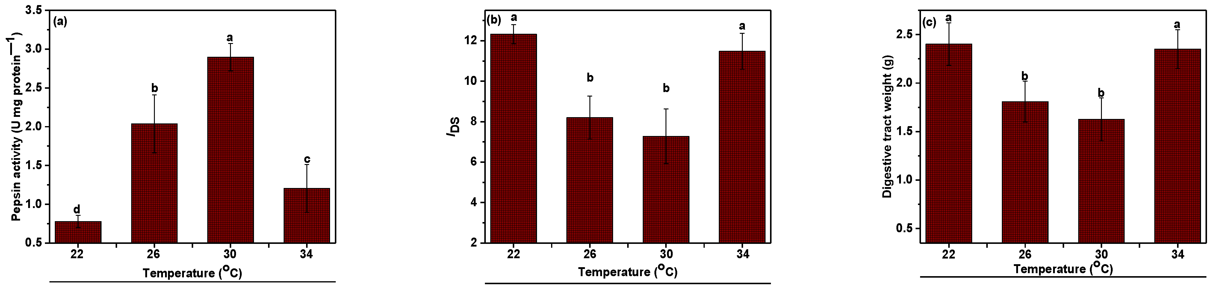
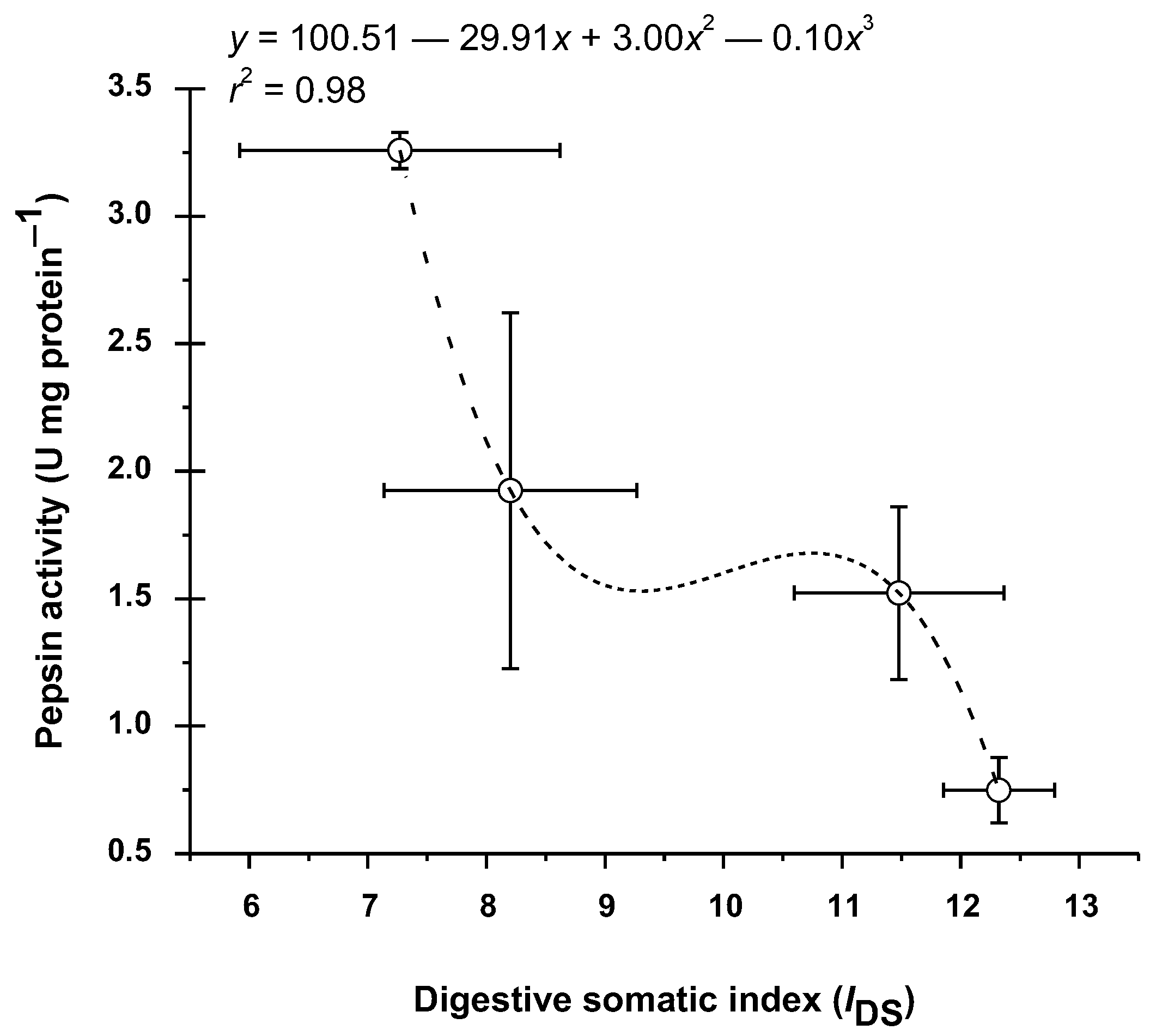
3.4. Pepsin Enzyme Activity
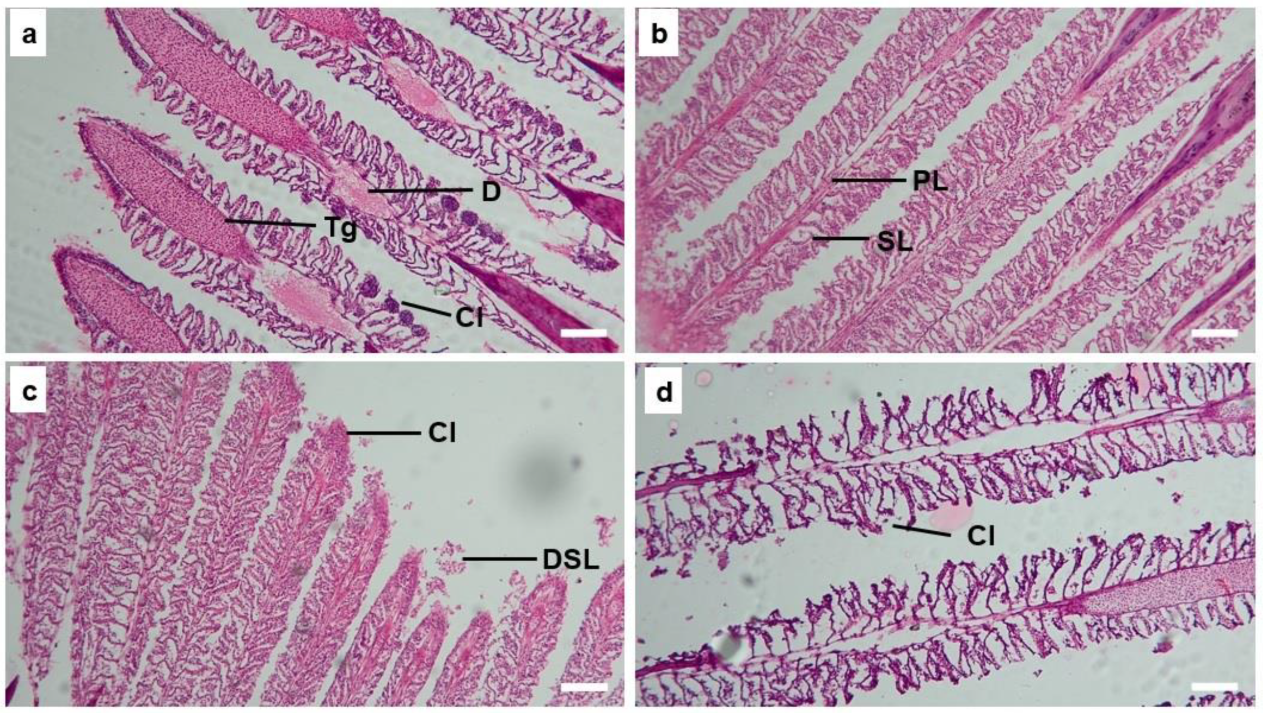
The relation between pepsin activity measured at four distinct exposure temperatures is displayed in Figure 5. Pepsin activities were measured at temperature profiles between 22 and 34 °C. Pepsin enzymatic activity showed a profile that increased from 22 °C to 30° and differed significantly (p < 0.05). In general, the ideal temperature for pepsin activity in the gut of B. gonionotus was observed at 30° (Figure 5). Fish fed commercial pellet diets showed an increased profile of pepsin enzymatic activity from 22 °C (0.78 ± 0.08 U mg protein−1) to 30 °C (2.90 ± 0.09 U mg protein−1) and differed significantly (p < 0.05). No significant effect was recorded at 22 and 34 °C (p > 0.05). Conversely, digestive tract weight showed an opposite trend of pepsin activity where the tract weight at 22 and 34 °C significantly increased from 26 and 30 °C (p < 0.05).
Figure 6 illustrates the correlation between the pepsin enzyme activity and the digestive somatic index (IDS) in the digestive tract of B. gonionotus. The link between IDS and pepsin enzyme activity was determined using a polynomial cubic model (r2 = 0.98), which showed a negative association between the two. Specifically, the temperature at which the pepsin activity was maximum was 30 °C. At the same temperature, the IDS value was lowest and the pepsin enzyme activities decreased as the IDS increased. Pepsin activity was at its lowest and the IDS value was at its highest at 22 °C (Figure 6).

3.5. Gill Histology

In-depth alterations in the gill tissues of fish subjected to various treatments were seen during the histological examinations (Figure 7, Table 3). At temperatures of 26 °C and 30 °C (Figure 7b,c), the gill tissues exhibited the typical well-formed primary and secondary lamellae. Primary lamellae (PL) are the initial structures involved in gas exchange, and secondary lamellae (SL) increase the surface area for more efficient gas exchange. The presence of well-formed lamellae indicates normal functioning and healthy gill tissues in these temperature groups. However, at temperatures of 22 °C and 34 °C (Figure 7a,d), the gill tissues showed distinct signs of damage. Branchitis is predominantly observed, combined with gill vascular lesions, and swelling in the secondary lamellar epithelium (Figure 7a,d). Hemorrhagic talengiactasis, consisting of extensive fibroplasia with histiocytic inflammatory cell infiltration, are dominant in secondary lamellae (Figure 7a,d). Clubbing which is the joining of gill filaments together has been seen in the primary and secondary gill lamellae (Figure 7a,c). In the base of the gill filament, swelling of the tissue and occasional disruption of the lamellae (broken or detachment) or degeneration is also present in primary and secondary gill lamellae (Figure 7b).

4. Discussion

As with all vertebrates, stress is the overall physiological reaction of fish to hazardous events [46]. This is a response to circumstances that are outside the ranges that are ideal for typical homeostatic responses, resulting in a disrupted state where regular regulatory adjustments are adequate. One of the environmental factors that has the most direct and significant impact on the numerous physiological processes of fish is water temperature. Each species of fish has a comfortable operating temperature range, and variations outside of this range cause stress and interfere with daily activity [47]. In the present study, we examined the effect of temperature on fish growth, hemato-biochemical responses, enzymatic activity, and gills structure of B. gonionotus. When the temperature was raised to the ideal level, fish performed better. Overall, the growth rate was highest in groups reared at 30 °C (Table 2). The experimental findings suggested that B. gonionotus can adapt to gradual increases in temperature. The fish did not suffer any adverse consequences, including irreparable damage, loss of balance, halting of breathing, or decreased food intake, while the temperature was maintained between 26 °C and 30 °C. This range of temperatures guaranteed the fish’s normal behavior and excellent survival rate. There was a significant decrease in food consumption when the water temperature was between 30 and 22 °C. The FCR values were notably lower in the group kept at 30 °C. When the temperature deviates from the optimal range, both FC decreases and FCR increases due to the impact on feed intake, food digestion time, and enzyme activity [14,48,49,50], and these findings might explain why we obtained satisfactory values for FC and FCR at 30 °C in our experiment. Our results were consistent with the observation for the Malabar blood snapper, which exhibited satisfactory FCR values at 26 and 30 °C [13,51]. Moreover, they noted that the FCE reached its maximum at 30 °C, indicating that the ideal temperature for FCE dropped with diet from 30 to 22 °C and from 30 to 34 °C. In Lates calcarifer, Katersky and Carter [52] demonstrated that SGR peaked between 33 and 36 °C, with no discernible change observed when the temperature was raised from 27 to 36 °C. When the temperature rose above 36 °C, they also discovered a decrease in growth efficiency. We observed that 26 to 30 °C is an appropriate temperature range for growth. The enhanced growth observed upon reaching the optimal temperature is consistent with earlier research conducted on Atlantic salmon [53]. Previous research has also documented a relationship between the growth rate and water temperature when there is free access to food [54,55]. Furthermore, the equilibrium between energy intake and expenditure determines the growth rate of fish, and temperature has an impact on both variables [56].
Fish’s hematological parameters are altered by stress, with subsequent effects on a variety of physiological systems [57,58,59]. According to the findings, most of the hematological parameters of B. gonionotus that were examined were significantly impacted by varying degrees of temperature fluctuations. The investigation’s findings demonstrated that B. gonionotus blood parameters were dramatically affected by low and high temperatures. The MCV increased while the RBC, HGB, PLT, HCT, MCH, and MCHC content significantly reduced. The values observed in this experiment are consistent with those found by Rhamdia and de Moraes [60] and Lermen et al. [61] who studied the hematologic characteristics of Rhamdia quelen. The crucian carp (Carassius carassius) treated at higher temperatures showed similar results [62]. The specific responses of fish to high temperature stress vary significantly depending on the species and the length of exposure as well as the species’ capacity for adaptation. Fish hematological profiles are altered by temperature fluctuations that are either above or below the ideal range which can result in hypoxia or anoxia [63,64]. According to Cech and Brauner [65], increased temperature reduces the solubility of oxygen in water, which results in oxygen deficiency in fish. According to Francis-Floyd et al. [66], fluctuating temperature also changes the dynamics of ammonia in the culture system in favor of unionized ammonia, which is poisonous and “causes stress and damages gills and other tissues, even in small amounts”. WBC level is a recognized indicator of fish health and is essential to enhance innate immunity or nonspecific immunity [22,67]. During the study, the WBC content in the current study considerably rose at the higher and lower temperature regimes (22 and 34 °C), potentially promoting antibody formation in response to a stressful environment.
Biochemical parameters of serum are considered as a dependable diagnostic tool for examining the health and nutritional condition of fish [68,69,70]. The serum biochemistry values differ among species and can be impacted by several biotic and abiotic elements, including food, age, sex of the fish, and water temperature [71]. The levels of alkaline phosphatase, albumin, globulin, A/G ratio, and total plasma protein in the B. gonionotus under study were comparable to those reported by Tavares-Dias et al. [72]. Although the data pertain to fish from tropical places where the temperature was steady throughout the year, these fluctuations may be linked to temperature changes that may have an impact on the biochemical blood levels [73]. The varying biochemical metabolism in different seasons may be the cause of the differences in the biochemical blood parameters [73,74]. Although the potential significance of serum protein as a fuel source for tissues during osmotic acclimation has not yet been explored, it might be associated with a metabolic reallocation of energy resources following the mobilization of carbohydrate storage. Fish cardiomyocytes and hepatocytes contain the majority of AST and ALT, which are key components of protein metabolism. Blood transaminase activity will rise because of the release of AST and ALT into the bloodstream caused by injury to or an increase in the permeability of liver and heart cells. Fish health status can thus be tracked using the serum AST and ALT activity [75]. The current study found that although there was a considerable variation in the AST and ALT values at different temperature levels, the values remained within the normal range, suggesting that the fish were in normal condition. To fill up the gaps in our understanding of this B. gonionotus, further research on a vast number of fish populations during various seasons, ages, sexes, and environmental conditions is necessary, as evidenced by the diversity in hematological and biochemical parameters found in this study.
For comparison research, figuring out the ideal temperature for enzyme activity could be interesting. Temperature changes typically have a significant impact on an enzyme’s ability to catalyze reactions. According to reports, many species’ maximum protease activity can occur in a wide temperature range, between 30 and 60 °C [76]. The surrounding temperature and the temperature inside the fish gut lumen are closely related. Thus, the temperature of the water has a variety of effects on fish digestion. The findings of the experiment examining the impact of temperature on pepsin enzyme activity indicated that pepsin functioned optimally at 30 °C. The temperature dropped to 22 °C, which caused a significant drop in enzyme activity. Turbot and redfish maintained almost half of the activity at low temperatures (5 °C). In contrast, seabream had less than 3.5 times this activity (p < 0.001) at the same temperature [77]. Since the structure of a protein is determined by its chemical bonds, the activity of the enzyme catalyzing a reaction would be affected or would decline [78]. When the temperature is raised from lower to higher, the enzyme activity rate is high because the more quickly molecules move, the more often substrates clash with the active sites on the enzyme [78]. The optimum pepsin activity at 30 °C in B. gonionotus gut reveals that the ideal temperature for the enzymes was higher than the environmental temperature; still, the findings from research on other fishes support this phenomenon [13,79,80]. Research has indicated that a drop in ambient temperature may have an adverse effect on fish’s capacity to hydrolyze food.
The gastrointestinal regions undergo morphological changes because of temperature adaptation. In colder and warmer weather, the IDS tended to be greater and recovered its initial levels when exposed to favorable conditions. Fish that have adapted to both cold and hot temperatures have larger guts, as seen by the increase in the unaltered cylinder surface area. These fish species have developed villi that are noticeably wider and taller than those of fish that have adjusted to the ideal temperature in the cold [81]. Comparable findings have also been described for Acipenser transmontanus, Acipenser naccarii, Oncorhynchus mykiss and Salmo salar L. 1758 [13,82,83,84]. The current findings demonstrated a relationship between stomach weight and development rate via absorption capacity. According to Bélanger et al. [85] and Mazumder et al. [13], the IDS of fish subjected to 26 and 30 °C was lower than that of fish exposed to lower and higher temperatures (22 and 34 °C). When fish species were fed less food, pepsin activity rose significantly in both low and high temperature exposure situations, exhibiting a tendency that was opposite to that of IDS. This is corroborated by an in-situ investigation conducted by Fu et al. [86], which found that from June to July, in A. japonicas pepsin-like activity increased considerably (fishes consuming less food or none at all). Moreover, Gao [87] discovered that deep estimation caused a surge in pepsin activity.
Fish gills are the key organs that are directly involved in metabolic processes [88,89]. The part of fish that comes into touch with water the most is the gill, which is sensitive to changes in the aquatic environment, including temperature. In addition to harming the gill organs, exposure to extreme temperatures impairs respiration [90,91]. In the present study, the histological examination revealed that B. gonionotus exposed to temperatures of 22 °C and 34 °C experienced significant alterations in their gill tissues, specifically in the form of splitting and shrinking lamellae. In contrast, fish exposed to 26 °C and 30 °C showed normal and healthy gill tissues with well-formed primary and secondary lamellae. Gill lamellae splitting and shrinking could negatively impact gas exchange efficiency and overall gill function [72]. The presence of well-formed lamellae suggests that the fish in these temperature groups are experiencing minimal stress, and their gills are efficiently performing their role in gas exchange as the fish gill is a multipurpose organ for aquatic gas exchange following Evans et al. [92]. At suboptimal temperatures of 22 °C, the gill tissues showed signs of lamellae splitting and shrinking. Lamellae splitting and shrinking can reduce the surface area available for gas exchange, potentially affecting the fish’s respiratory efficiency. On the other hand, at a higher temperature of 34 °C, similar signs of lamellae splitting and shrinking were observed. Amir et al. [90] also subjected hybrid catfish to three different temperatures and observed skin darkening in fish acclimated with increasing temperatures. They discovered that fish acclimated to 37 °C had thicker epithelial layers, and they recommended maintaining the ideal temperature in the aquaculture environment to avoid physiological harm to the fish as well as decreased growth and productivity. These results have ramifications for our knowledge of fish physiological responses to temperature changes. They highlight the importance of maintaining suitable temperature conditions in aquatic environments to ensure the well-being and optimal functioning of fish gills, which are critical for their survival and adaptation to changing environmental conditions. This finding is also supported by Saber [93] who investigated the gill tissues of common carp Cyprinus carpio L. where gill sections of this fish species showed marked histological lesions during low and high temperatures, and by Zaman et al. [94] who investigated liver and kidney of Puntias sarana and found numerous histopathological changes.

5. Conclusions

The main findings of this study focused on the growth performances, hemo-biochemical reactions, enzymatic activity, and gills structural alterations of B. gonionotus under diverse temperature ranges encountered in natural conditions. The results of this study show that B. gonionotus should be cultured at 30 °C since this temperature promotes the enhanced enzymatic activity that results in a quicker digesting process and, eventually, a faster growth rate of this economically significant fish species. Changes in blood characteristics are significant indicators in tracking the management of fish physiology, as shown by differences in hemo-biochemical parameters because of various temperature management strategies. These results suggest that temperature plays a crucial role in influencing the structural integrity and function of gill tissues in B. gonionotus. A quantitative histological study revealed that fish exposed to 26 °C and 30 °C showed normal and healthy gill tissues with well-formed primary and secondary lamellae. We can draw the conclusion that the information at hand can help to explain how B. gonionotus is able to withstand a wide range of ambient temperatures.

Author Contributions

Conceptualization, Methodology, Formal analysis: S.K.M. and S.K.D. Investigation and writing original draft: S.D. Writing—review & editing: S.K.D., M.S.A., M.L.R., M.A.A.M., S.M.I.K. and D.P. Project administration and supervision: S.K.M. and M.A.S. Funding acquisition: S.K.M. All authors have read and agreed to the published version of the manuscript.

Funding

This research was funded by the Ministry of Science and Technology, Bangladesh under the Special Allocation for Science and Technology fund grant (Grant no. BS-2021-196).

Data Availability Statement

The data presented in this study are available on request from the corresponding author.

Conflicts of Interest

The authors declare no conflict of interest.

References

  1. Cline, A.J.; Hamilton, S.L.; Logan, C.A. Effects of multiple climate change stressors on gene expression in blue rockfish (Sebastes mystinus). Comp. Biochem. Physiol. Part A Mol. Integr. Physiol. 2020, 239, 110580. [Google Scholar] [CrossRef] [PubMed]
  2. O’Gorman, E.J.; Ólafsson, Ó.P.; Demars, B.O.; Friberg, N.; Guðbergsson, G.; Hannesdóttir, E.R.; Jackson, M.C.; Johansson, L.S.; McLaughlin, Ó.B.; Ólafsson, J.S.; et al. Temperature effects on fish production across a natural thermal gradient. Glob. Chang. Biol. 2016, 22, 3206–3220. [Google Scholar] [CrossRef] [PubMed]
  3. Jensen, L.B.; Boltana, S.; Obach, A.; McGurk, C.; Waagbø, R.; MacKenzie, S. Investigating the underlying mechanisms of temperature-related skin diseases in Atlantic salmon, Salmo salar L., as measured by quantitative histology, skin transcriptomics and composition. J. Fish Dis. 2015, 38, 977–992. [Google Scholar] [CrossRef] [PubMed]
  4. Pfahl, S.; O’Gorman, P.A.; Fischer, E.M. Understanding the regional pattern of projected future changes in extreme precipitation. Nat. Clim. Chang. 2017, 7, 423–427. [Google Scholar] [CrossRef]
  5. Webster, P.J. Dynamics of the Tropical Atmosphere and Oceans; John Wiley & Sons: Hoboken, NJ, USA, 2020. [Google Scholar]
  6. Islam, S.M.; Sultana, R.; Imran, M.; Jannat, M.F.T.; Ashaf-Ud-Doulah, M.; Rohani, M.F.; Brown, C.; Shahjahan, M. Elevated temperature affects growth and hemato-biochemical parameters, inducing morphological abnormalities of erythrocytes in Nile tilapia Oreochromis niloticus. Aquac. Res. 2020, 51, 4361–4371. [Google Scholar] [CrossRef]
  7. Yilmaz, H.A.; Turkmen, S.; Kumlu, M.; Eroldogan, O.T.; Perker, N. Alteration of growth and temperature tolerance of European sea bass (Dicentrarchus labrax linnaeus 1758) in different temperature and salinity combinations. Turk. J. Fish. Aquat. Sci. 2020, 20, 331–340. [Google Scholar] [CrossRef]
  8. Nadermann, N.; Seward, R.K.; Volkoff, H. Effects of potential climate change-induced environmental modifications on food intake and the expression of appetite regulators in goldfish. Comp. Biochem. Physiol. Part A Mol. Integr. Physiol. 2019, 235, 138–147. [Google Scholar] [CrossRef]
  9. Burgerhout, E.; Lokman, P.M.; van den Thillart, G.E.; Dirks, R.P. The time-keeping hormone melatonin: A possible key cue for puberty in freshwater eels (Anguilla spp.). Rev. Fish Biol. Fish. 2019, 29, 1–21. [Google Scholar] [CrossRef]
  10. Cheng, S.Y.; Chen, C.S.; Chen, J.C. Salinity and temperature tolerance of brown-marbled grouper Epinephelus fuscoguttatus. Fish Physiol. Biochem. 2013, 39, 277–286. [Google Scholar] [CrossRef]
  11. Shahjahan, M.; Kitahashi, T.; Ando, H. Temperature affects sexual maturation through the control of kisspeptin, kisspeptin receptor, GnRH and GTH subunit gene expression in the grass puffer during the spawning season. Gen. Comp. Endocrinol. 2017, 243, 138–145. [Google Scholar] [CrossRef]
  12. Slater, M.; D’Abramo, L.; Engle, C.R. Aquaculture research priorities for the next decade: A global perspective. J. World Aquac. Soc. 2018, 49, 3–6. [Google Scholar] [CrossRef]
  13. Mazumder, S.K.; Das, S.K.; Rahim, S.M.; Abd Ghaffar, M. Temperature and diet effect on the pepsin enzyme activities, digestive somatic index and relative gut length of Malabar blood snapper (Lutjanus malabaricus Bloch & Schneider, 1801). Aquac. Rep. 2018, 9, 1–9. [Google Scholar]
  14. Mazumder, S.K.; Fivelstad, S.; Ghaffar, M.A.; Das, S.K. Haematological and Biochemical Responses of Juvenile Malabar Blood Snapper (Lutjanus malabaricus Bloch & Schneider, 1801) Exposed to Different Rearing Temperatures and Diets. Sains Malays. 2019, 48, 1790–1810. [Google Scholar]
  15. Pepin, P. Effect of temperature and size on development, mortality, and survival rates of the pelagic early life history stages of marine fish. Can. J. Fish. Aquat. Sci. 1991, 48, 503–518. [Google Scholar] [CrossRef]
  16. Fu, K.K.; Fu, C.; Qin, Y.L.; Bai, Y.; Fu, S.J. The thermal acclimation rate varied among physiological functions and temperature regimes in a common cyprinid fish. Aquaculture 2018, 495, 393–401. [Google Scholar] [CrossRef]
  17. Maulvault, A.L.; Barbosa, V.; Alves, R.; Custódio, A.; Anacleto, P.; Repolho, T.; Ferreira, P.P.; Rosa, R.; Marques, A.; Diniz, M. Ecophysiological responses of juvenile seabass (Dicentrarchus labrax) exposed to increased temperature and dietary methylmercury. Sci. Total Environ. 2017, 586, 551–558. [Google Scholar] [CrossRef] [PubMed]
  18. Morgan, J.D.; Iwama, G.K. Energy cost of NaCl transport in isolated gills of cutthroat trout. Am. J. Physiol.-Regul. Integrat. Comp. Physiol. 1999, 277, R631–R639. [Google Scholar] [CrossRef]
  19. Adams, S.M. Status and use of biological indicators for evaluating the effects of stress on fish. [Formerly used by DOE/TIC for titles for which valid CODEN was not available. Now invalid] 92-DEC-15; (Country unknown/Code not available). 1990, 8.
  20. Ruane, N.M.; Nolan, D.T.; Rotllant, J.; Costelloe, J.; Bonga, S.W. Experimental exposure of rainbow trout Oncorhynchus mykiss (Walbaum) to the infective stages of the sea louse Lepeophtheirus salmonis (Krøyer) influences the physiological response to an acute stressor. Fish Shellfish Immunol. 2000, 10, 451–463. [Google Scholar] [CrossRef]
  21. Sharmin, S.; Shahjahan, M.; Hossain, M.A.; Haque, M.A.; Rashid, H. Histopathological changes in liver and kidney of common carp exposed to sub-lethal doses of malathion. Pak. J. Zool. 2015, 47, 1495–1498. [Google Scholar]
  22. Islam, S.M.; Rahman, M.A.; Nahar, S.; Uddin, M.H.; Haque, M.M.; Shahjahan, M. Acute toxicity of an organophosphate insecticide sumithion to striped catfish Pangasianodon hypophthalmus. Toxicol. Rep. 2019, 6, 957–962. [Google Scholar] [CrossRef] [PubMed]
  23. Jaffer, N.S.; Rabee, A.M.; Al-Chalabi, S.M. Biochemical and hematological parameters and histological alterations in fish Cyprinus carpio L. as biomarkers for water pollution with chlorpyrifos. Hum. Ecol. Risk Assess. Int. J. 2017, 23, 605–616.S. [Google Scholar] [CrossRef]
  24. Nimet, J.; Neves, M.P.; Viana, N.P.; de Arruda Amorim, J.P.; Delariva, R.L. Histopathological alterations in gills of a fish (Astyanax bifasciatus) in neotropical streams: Negative effects of riparian forest reduction and presence of pesticides. Environ. Monit. Assess. 2020, 192, 58. [Google Scholar] [CrossRef] [PubMed]
  25. Rahman, M.M.; Hossain, M.Y.; Ahamed, F.; Fatematuzzhura, S.B.; Abdallah, E.M.; Ohtomi, J. Biodiversity in the Padma distributary of the Ganges River, Northwestern Bangladesh: Recommendations for conservation. World J. Zool. 2012, 7, 328–337. [Google Scholar]
  26. Rahman, M.Z.; Zaman, M.; Hossain, A.; Akter, S.; Rahman, M.M.; Mamun, M.A.A.; Alam, M.M.M.; Khalil, S.M.I. Ovarian Histology and Histopathology of Olive Barb, Puntius sarana Exposed to Endocrine Disrupting Chemical (17-A Methyl Testosterone) in Laboratory Condition. Aquac. Stud. 2023, 24, AQUAST1177. [Google Scholar] [CrossRef]
  27. Mondal, D.; Rahman, T.; Paul, S.; Islam, M.; Miah, I. Effects of probiotic supplementation on the growth performance of Thai silver barb (Barbonymus gonionotus) (bleeker, 1850) fry. Fundam. Appl. Agric. 2019, 4, 950–958. [Google Scholar] [CrossRef]
  28. Mohanta, K.N.; Mohanty, S.N.; Jena, J.K.; Sahu, N.P. Apparent protein, lipid and energy digestibility coefficients of some commonly used feed ingredients in formulated pelleted diets for silver barb, Puntius gonionotus. Aquac. Nutr. 2006, 12, 211–218. [Google Scholar] [CrossRef]
  29. Mostakim, G.M.; Zahangir, M.M.; Mishu, M.M.; Rahman, M.K.; Islam, M.S. Alteration of blood parameters and histoarchitecture of liver and kidney of silver barb after chronic exposure to quinalphos. J. Toxicol. 2015, 2015, 415984. [Google Scholar] [CrossRef]
  30. Sadiqul, I.M.; Ferdous, Z.; Nannu, M.T.A.; Mostakim, G.M.; Rahman, M.K. Acute exposure to a quinalphos containing insecticide (convoy) causes genetic damage and nuclear changes in peripheral erythrocytes of silver barb, Barbonymus gonionotus. Environ. Pollut. 2016, 219, 949–956. [Google Scholar] [CrossRef]
  31. Nayak, M.; Saha, A.; Pradhan, A.; Samanta, M.; Giri, S.S. Dietary fish oil replacement by linseed oil: Effect on growth, nutrient utilization, tissue fatty acid composition and desaturase gene expression in silver barb (Puntius gonionotus) fingerlings. Comp. Biochem. Physiol. Part B Biochem. Mol. Biol. 2017, 205, 1–12. [Google Scholar] [CrossRef]
  32. Jahan, A.; Nipa, T.T.; Islam, S.M.; Uddin, M.H.; Islam, M.S.; Shahjahan, M. Striped catfish (Pangasianodon hypophthalmus) could be suitable for coastal aquaculture. J. Appl. Ichthyol. 2019, 35, 994–1003. [Google Scholar] [CrossRef]
  33. Wangsongsak, A.; Utarnpongsa, S.; Kruatrachue, M.; Ponglikitmongkol, M.; Pokethitiyook, P.; Sumranwanich, T. Alterations of organ histopathology and metallolhionein mRNA expression in silver barb, Puntius gonionotus during subchronic cadmium exposure. J. Environ. Sci. 2007, 19, 1341–1348. [Google Scholar] [CrossRef] [PubMed]
  34. Romano, N.; Ashikin, M.; Teh, J.C.; Syukri, F.; Karami, A. Effects of pristine polyvinyl chloride fragments on whole body histology and protease activity in silver barb Barbodes gonionotus fry. Environ. Pollut. 2018, 237, 1106–1111. [Google Scholar] [CrossRef] [PubMed]
  35. Shah, S.L.; Altindag, A. Haematological parameters of tench (Tinca tinca L., 1758) on acute and chronic exposures of lethal and sublethal treatments of cadmium. Fresenius Environ. Bull. 2004, 13, 1477–1481. [Google Scholar]
  36. Blaxhall, P.C.; Daisley, K.W. Routine haematological methods for use with fish blood. J. Fish Biol. 1973, 5, 771–781. [Google Scholar] [CrossRef]
  37. Shah, S.L.; Altindağ, A. Alterations in the immunological parameters of Tench (Tinca tinca L. 1758) after acute and chronic exposure to lethal and sublethal treatments with mercury, cadmium and lead. Turk. J. Vet. Anim. Sci. 2005, 29, 1163–1168. [Google Scholar]
  38. Wright, G.; Sanderson, J.M. Brain damage and mortality in dogs following pulsatile and non-pulsatile blood flows in extracorporeal circulation. Thorax 1972, 27, 738–747. [Google Scholar] [CrossRef]
  39. Ye, H.; Xu, M.; Liu, Q.; Sun, Z.; Zou, C.; Chen, L.; Su, N.; Ye, C. Effects of replacing fish meal with soybean meal on growth performance, feed utilization and physiological status of juvenile obscure puffer, Takifugu obscurus. Comp. Biochem. Physiol. Part C Toxicol. Pharmacol. 2019, 216, 75–81. [Google Scholar] [CrossRef]
  40. Abolfathi, M.; Hajimoradloo, A.; Ghorbani, R.; Zamani, A. Compensatory growth in juvenile roach Rutilus caspicus: Effect of starvation and re-feeding on growth and digestive surface area. J. Fish. Biol. 2012, 81, 1880–1890. [Google Scholar] [CrossRef]
  41. Bradford, M.M. A rapid and sensitive method for the quantitation of microgram quantities of protein utilizing the principle of protein-dye binding. Anal. Biochem. 1976, 72, 248–254. [Google Scholar] [CrossRef]
  42. Natalia, Y.; Hashim, R.; Ali, A.; Chong, A. Characterization of digestive enzymes in a carnivorous ornamental fish, the Asian bony tongue Scleropages formosus (Osteoglossidae). Aquaculture 2004, 233, 305–320. [Google Scholar] [CrossRef]
  43. Anson, M.L. The estimation of pepsin, trypsin, papain, and cathepsin with hemoglobin. J. Gen. Physiol. 1938, 22, 79–89. [Google Scholar] [CrossRef] [PubMed]
  44. Huggett, R.J. Biomarkers: Biochemical, Physiological, and Histological Markers of Anthropogenic Stress; CRC Press: Boca Raton, FL, USA, 2018. [Google Scholar]
  45. Mašković, J.M.; Marković, G.; Mašković, P.Z. Analytical and environmental control of water quality of the Bjelica river. Acta Agric. Serbica 2018, 23, 111–120. [Google Scholar] [CrossRef]
  46. Schreck, C.B.; Tort, L. The concept of stress in fish. In Fish Physiology; Academic Press: Cambridge, MA, USA, 2016; Volume 35, pp. 1–34. [Google Scholar]
  47. Beitinger, T.L.; Bennett, W.A. Quantification of the role of acclimation temperature in temperature tolerance of fishes. Environ. Biol. Fish. 2000, 58, 277–288. [Google Scholar] [CrossRef]
  48. Mazumder, S.K.; Ghaffar, M.A.; Das, S.K. Exploring the suitable temperature and diet for growth and gastric emptying time of juvenile Malabar Blood Snapper (Lutjanus malabaricus Bloch & Schneider, 1801). Thalass. Int. J. Mar. Sci. 2019, 35, 29–41. [Google Scholar]
  49. Jun, Q.; Pao, X.; Haizhen, W.; Ruiwei, L.; Hui, W. Combined effect of temperature, salinity and density on the growth and feed utilization of Nile tilapia juveniles (Oreochromis niloticus). Aquac. Res. 2012, 43, 1344–1356. [Google Scholar] [CrossRef]
  50. Handeland, S.O.; Imsland, A.K.; Stefansson, S.O. The effect of temperature and fish size on growth, feed intake, food conversion efficiency and stomach evacuation rate of Atlantic salmon post-smolts. Aquaculture 2008, 283, 36–42. [Google Scholar] [CrossRef]
  51. Mazumder, S.K.; Das, S.K.; Bakar, Y.; Ghaffar, M.A. Effects of temperature and diet on length-weight relationship and condition factor of the juvenile Malabar blood snapper (Lutjanus malabaricus Bloch & Schneider, 1801). J. Zhejiang Univ. Sci. 2016, 17, 580–590. [Google Scholar]
  52. Katersky, R.S.; Carter, C.G. Growth efficiency of juvenile barramundi, Lates calcarifer, at high temperatures. Aquaculture 2005, 250, 775–780. [Google Scholar] [CrossRef]
  53. Handeland, S.; Arnesen, A.; Stefansson, S. Seawater adaptation and growth of post-smolt Atlantic salmon (Salmo salar) of wild and farmed strains. Aquaculture 2003, 220, 367–384. [Google Scholar] [CrossRef]
  54. Dong, Y.; Dong, S. Growth and oxygen consumption of the juvenile sea cucumber Apostichopus japonicus (Selenka) at constant and fluctuating water temperatures. Aquac. Res. 2006, 37, 1327–1333. [Google Scholar] [CrossRef]
  55. Jobling, M.; Meløy, O.; Dos Santos, J.; Christiansen, B. The compensatory growth response of the Atlantic cod: Effects of nutritional history. Aquac. Int. 1994, 2, 75–90. [Google Scholar] [CrossRef]
  56. Diana, J.S. Biology and Ecology of Fishes; John Wiley & Sons, Inc.: Hoboken, NJ, USA, 1995. [Google Scholar]
  57. Ferreira, F.; Santos, M.M.; Reis-Henriques, M.A.; Vieira, M.N.; Monteiro, N.M. The annual cycle of spermatogenesis in Lipophrys pholis (Blenniidae), a recently proposed sentinel species for pollution monitoring. Ichthyol. Res. 2011, 58, 360–365. [Google Scholar] [CrossRef]
  58. Islam, M.J.; Kunzmann, A.; Thiele, R.; Slater, M.J. Effects of extreme ambient temperature in European seabass, Dicentrarchus labrax acclimated at different salinities: Growth performance, metabolic and molecular stress responses. Sci. Total Environ. 2020, 735, 139371. [Google Scholar] [CrossRef] [PubMed]
  59. Shahjahan, M.; Uddin, M.H.; Bain, V.; Haque, M.M. Increased water temperature altered hemato-biochemical parameters and structure of peripheral erythrocytes in striped catfish Pangasianodon hypophthalmus. Fish Physiol. Biochem. 2018, 44, 1309–1318. [Google Scholar] [CrossRef] [PubMed]
  60. Rhamdia, P.O.J.; de Moraes, G.M.F.R. Características hematológicas de teleósteos brasileiros. IV. Variáveis do jundiá Rhamdia quelen (Pimelodidae). Ciência Rural 2002, 32, 693–698. [Google Scholar]
  61. Lermen, C.L.; Lappe, R.; Crestani, M.; Vieira, V.P.; Gioda, C.R.; Schetinger, M.R.C.; Baldisserotto, B.; Moraes, G.; Morsch, V.M. Effect of different temperature regimes on metabolic and blood parameters of silver catfish Rhamdia quelen. Aquaculture 2004, 239, 497–507. [Google Scholar] [CrossRef]
  62. Sikorska, J.; Kondera, E.; Kamiński, R.; Ługowska, K.; Witeska, M.; Wolnicki, J. Effect of four rearing water temperatures on some performance parameters of larval and juvenile crucian carp, Carassius carassius, under controlled conditions. Aquac. Res. 2018, 49, 3874–3880. [Google Scholar] [CrossRef]
  63. Carvalho, C.S.; Fernandes, M.N. Effect of temperature on copper toxicity and hematological responses in the neotropical fish Prochilodus scrofa at low and high pH. Aquaculture 2006, 251, 109–117. [Google Scholar] [CrossRef]
  64. Hedayati, A.; Tarkhani, R. Hematological and gill histopathological changes in iridescent shark, Pangasius hypophthalmus (Sauvage, 1878) exposed to sublethal diazinon and deltamethrin concentrations. Fish Physiol. Biochem. 2014, 40, 715–720. [Google Scholar] [CrossRef]
  65. Cech, J.J.; Brauner, C.J. Techniques in whole animal respiratory physiology. Encycl. Fish Physiol. Genome Environ. 2011, 2, 846–853. [Google Scholar]
  66. Francis-Floyd, R.; Watson, C.; Petty, D.; Pouder, D.B. Ammonia in Aquatic Systems; UF/IFAS University of Florida (UF)/Institute of Food and Agricultural Sciences (IFAS), FA: Gainesville, FL, USA, 2009; Volume 16, pp. 1–4. [Google Scholar]
  67. Roberts, R.J. Fish Pathology; John Wiley & Sons: Hoboken, NJ, USA, 2012. [Google Scholar]
  68. Satheeshkumar, P.; Ananthan, G.; Kumar, D.S.; Jagadeesan, L. Haematology and biochemical parameters of different feeding behaviour of teleost fishes from Vellar estuary, India. Comp. Clin. Pathol. 2012, 21, 1187–1191. [Google Scholar] [CrossRef]
  69. Rashmeei, M.; Shekarabi, S.P.H.; Mehrgan, M.S.; Paknejad, H. Stimulatory effect of dietary chasteberry (Vitex agnus-castus) extract on immunity, some immune-related gene expression, and resistance against Aeromonas hydrophila infection in goldfish (Carassius auratus). Fish Shellfish Immunol. 2020, 107, 129–136. [Google Scholar] [CrossRef] [PubMed]
  70. Gheytasi, A.; Hosseini Shekarabi, S.P.; Islami, H.R.; Mehrgan, M.S. Feeding rainbow trout, Oncorhynchus mykiss, with lemon essential oil loaded in chitosan nanoparticles: Effect on growth performance, serum hemato-immunological parameters, and body composition. Aquac. Int. 2021, 29, 2207–2221. [Google Scholar] [CrossRef]
  71. Jawad, L.A.; Al-Mukhtar, M.A.; Ahmed, H.K. The relationship between haematocrit and some biological parameters of the Indian shad, Tenualosa ilisha (Family Clupeidae). Anim. Biodivers. Conserv. 2004, 27, 47–52. [Google Scholar]
  72. Tavares-Dias, M.; Frascá-Scorvo, C.; Novato, P.; Moraes, F. Hematological characteristics of hybrid Florida red tilapia, Oreochromis urolepis hornorun x O. mossambicus under intensive rearing. In Proceedings of the International Symposium on Tilapia Aquaculture, Rio de Janeiro, Brazil, 3–7 September 2000; pp. 533–541. [Google Scholar]
  73. Adeyemo, O.K.; Agbede, S.A.; Olaniyan, A.O.; Shoaga, O.A. The haematological response of Clarias gariepinus to changes in acclimation temperature. Afr. J. Biomed. Res. 2003, 6, 105–108. [Google Scholar] [CrossRef]
  74. Chavin, W.; Young, J.E. Factors in the determination of normal serum glucose levels of goldfish, Carassius auratus L. Comp. Biochem. Phys. 1970, 33, 629–653. [Google Scholar] [CrossRef]
  75. Yuan, W.; Li, X.; Kangjian, Y. Effect beta-cypermethrin on GPT and GOT activities of crucian serum. Nongye Kexue Yu Jishu 2005, 6, 20–23. [Google Scholar]
  76. Chen, M.; Zhang, X.; Gao, T.; Chen, C. Effects of temperature, pH and NaCl on protease activity in digestive tract of young turbot, Scophthalmus maximus. Chin. J. Oceanol. Limnol. 2006, 24, 300–306. [Google Scholar]
  77. Munilla-Morán, R.; Saborido-Rey, F. Digestive enzymes in marine species. I. Proteinase activities in gut from redfish (Sebastes mentella), seabream (Sparus aurata) and turbot (Scophthalmus maximus). Comp. Biochem. Physiol. Part B Biochem. Mol. Biol. 1996, 113, 395–402. [Google Scholar] [CrossRef]
  78. Reece, S.Y.; Hamel, J.A.; Sung, K.; Jarvi, T.D.; Esswein, A.J.; Pijpers, J.J.; Nocera, D.G. Wireless solar water splitting using silicon-based semiconductors and earth-abundant catalysts. Science 2011, 334, 645–648. [Google Scholar] [CrossRef] [PubMed]
  79. Lazo, J.P.; Mendoza, R.; Holt, G.J.; Aguilera, C.; Arnold, C.R. Characterization of digestive enzymes during larval development of red drum (Sciaenops ocellatus). Aquaculture 2007, 265, 194–205. [Google Scholar] [CrossRef]
  80. Xiong, D.M.; Xie, C.X.; Zhang, H.J.; Liu, H.P. Digestive enzymes along digestive tract of a carnivorous fish Glyptosternum maculatum (Sisoridae, Siluriformes). J. Anim. Physiol. Anim. Nutr. 2011, 95, 56–64. [Google Scholar] [CrossRef] [PubMed]
  81. Lee, J.A.; Cossins, A.R. Adaptation of intestinal morphology in the temperature-acclimated carp, Cyprinus carpio L. Cell Tissue Res. 1988, 251, 451–456. [Google Scholar] [CrossRef] [PubMed]
  82. Furné, M.; García-Gallego, M.; Hidalgo, M.C.; Morales, A.E.; Domezain, A.; Domezain, J.; Sanz, A. Effect of starvation and refeeding on digestive enzyme activities in sturgeon (Acipenser naccarii) and trout (Oncorhynchus mykiss). Comp. Biochem. Physiol. Part A Mol. Integr. Physiol. 2008, 149, 420–425. [Google Scholar] [CrossRef] [PubMed]
  83. Krogdahl, Å.; Bakke-McKellep, A.M. Fasting and refeeding cause rapid changes in intestinal tissue mass and digestive enzyme capacities of Atlantic salmon (Salmo salar L.). Comp. Biochem. Physiol. Part A Mol. Integr. Physiol. 2005, 141, 450–460. [Google Scholar] [CrossRef] [PubMed]
  84. Storebakken, T.; Shearer, K.D.; Refstie, S.; Lagocki, S.; McCool, J. Interactions between salinity, dietary carbohydrate source and carbohydrate concentration on the digestibility of macronutrients and energy in rainbow trout (Oncorhynchus mykiss). Aquaculture 1998, 163, 347–359. [Google Scholar] [CrossRef]
  85. Bélanger, F.; Blier, P.U.; Dutil, J.D. Digestive capacity and compensatory growth in Atlantic cod (Gadus morhua). Fish Physiol. Biochem. 2002, 26, 121–128. [Google Scholar] [CrossRef]
  86. Fu, X.; Xue, C.; Miao, B.; Li, Z.; Gao, X.; Hirata, T. Distribution and seasonal activity variation of proteases in digestive tract of sea cucumber Stichopus japonicus. Fish. Sci. 2006, 72, 1130–1132. [Google Scholar] [CrossRef]
  87. Gao, F.; Yang, H.; Xu, Q.; Wang, F.; Liu, G. Effect of water temperature on digestive enzyme activity and gut mass in sea cucumber Apostichopus japonicus (Selenka), with special reference to aestivation. Chin. J. Oceanol. Limnol. 2009, 27, 714–722. [Google Scholar] [CrossRef]
  88. Islam, M.R.; Hossain, M.A.; Afrose, F.; Roy, N.C.; Iqbal, M.M. Effect of temperature on the growth performance, haematological properties and histomorphology of gill, intestine and liver tissues in juvenile butter catfish Ompok bimaculatus. Aquac. Fish Fish. 2022, 2, 277–286. [Google Scholar] [CrossRef]
  89. Maftuch, M.; Sanoesi, E.; Farichin, I.; Saputra, B.A.; Ramdhani, L.; Hidayati, S.; Fitriyah, N.; Prihanto, A.A. Histopathology of gill, muscle, intestine, kidney, and liver on Myxobolus sp.-infected Koi carp (Cyprinus carpio). J. Parasit. Dis. 2018, 42, 137–143. [Google Scholar] [CrossRef] [PubMed]
  90. Amir, F.; Muchlisin, Z.A.; Nur, F.M.; Fadli, N.; Siti-Azizah, M.N.; Wilkes, M.; Tang, U.; Hasan, B.; Batubara, A.S.; Kocabas, F.K.; et al. Effect of increasing water temperature on the physiology and gill histology of Barramundi, Lates calcarifer (Pisces, Perciformes) fingerlings. Int. Aquat. Res. 2022, 14, 263–273. [Google Scholar]
  91. Flores-Lopes, F.; Thomaz, A.T. Histopathologic alterations observed in fish gills as a tool in environmental monitoring. Braz. J. Biol. 2011, 71, 179–188. [Google Scholar] [CrossRef]
  92. Evans, D.H.; Piermarini, P.M.; Choe, K.P. The multifunctional fish gill: Dominant site of gas exchange, osmoregulation, acid-base regulation, and excretion of nitrogenous waste. Physiol. Rev. 2005, 85, 97–177. [Google Scholar] [CrossRef]
  93. Saber, T.H. Histological adaptation to thermal changes in gills of common carp fishes Cyprinus carpio L. Rafidain J. Sci. 2011, 22, 46–55. [Google Scholar] [CrossRef]
  94. Zaman, M.; Khalil, S.M.I.; Rahman, M.Z.; Hossain, A.; Al Mamun, M.A.; Rahman, M.M.; Alam, M.M. Evaluation of Histopathological Alterations in the Liver and Kidney of Olive Barb (Puntius sarana, Hamilton 1822) as an Indicator of the Surma River’s Pollution. Aquat. Sci. Eng. 2023, 38, 81–88. [Google Scholar] [CrossRef]
Figure 1. Protocol for the experiment. There were four treatments in the experiment, and the fish were kept for 60 days each.
Water 16 00292 g001
Figure 2. Changes in the mean (a) total length and (b) body weight of B. gonionotus reared at four temperatures over 60 days. The means values shared with different letters differ significantly (p < 0.05).
Water 16 00292 g002
Figure 3. Ematological changes in B. gonionotus exposed to four different temperatures (22, 26, 30, 34 °C) (a) red blood cell, (b) white blood cell, (c) haemoglobin, (d) platelets, (e) mean corpuscular haemoglobin, (f) mean corpuscular haemoglobin concentration, (g) hematocrit, (h) mcv, (i) mean platelet volume. Within the same subfigure, the mean values in bars indicated by different letters are significantly different (p < 0.05).
Water 16 00292 g003
Figure 4. Effects of temperature fluctuations on the biochemical responses of B. gonionotus reared for 60 days (a) glucose, (b) triglycerides, (c) cholesterol, (d) total protein, (e) serum albumin, (f) HCO3 (TCO3), (g) Na+, (h) K+, (i) Cl, (j) AP, (k) AST, (l) serum creatinine, (m) bilirubin, (n) uric acid, (o) Ca+, (p) HDL, (q) LDL, (r) HDL/LDL ratio. Within the same subfigure, the bars indicated by different letters are significantly different (p < 0.05).
Water 16 00292 g004aWater 16 00292 g004b
Figure 5. Pepsin enzymatic activities of B. gonionotus at different temperatures (a) pepsin activity, (b) IDS, (c) digestive tract weight. Within the same subfigure, the bars with different letters are significantly different (p < 0.05).
Water 16 00292 g005
Figure 6. Correlation between digestive somatic index and specific pepsin activity in the gut contents of B. gonionotus at different temperatures. Values are means ± SE.
Water 16 00292 g006
Figure 7. Comparative microphotographs indicating the differences in the structures of gills lamellae of B. gonionotus acclimated to different temperatures ((a) 22 °C; (b) 26 °C; (c) 30 °C; (d) 34 °C). (a) consists of numerous talengiactasis (Tg) with hyperplasia and hypertrophy in the primary and secondary gill lamellae; clubbing (Cl) and detachment of in the SL (DSL); (b) consists of regular and symmetrical structure PL and SL with few abnormalities like occasional hemorrhage in base of PL and talengiactasis in the top of PL; (c) detachment of the PL from the base and SL, occasional clubbing and hemorrhage have been observed and (d) loss of normal structure, clubbing and talengiactesis were frequent. Scale bar: 200 µm; Stain: H & E.
Water 16 00292 g007
Table 1. Water quality parameters recorded throughout the experiment.
Parameters22 °C26 °C30 °C34 °C
Temperature (°C)22 ± 0.8426 ± 0.6230 ± 0.3834 ± 0.61
* TH (mg L−1)123.33 ± 17.33110.36 ± 24.36118.29 ± 21.37112.16 ± 16.21
* NH3-N (mg L−1)0.24 ± 0.0540.16 ± 0.0180.31 ± 0.0240.19 ± 0.031
pH6.91 ± 0.067.51 ± 0.247.66 ± 0.637.81 ± 0.96
Note: * TH: total hardness and NH3-N: ammoniacal nitrogen.
Table 2. Growth response of B. gonionotus at different temperatures for 60 days.
VariablesTemperature (°C)
22263034
Initial total length (cm)11.13 ± 1.00 a11.45 ± 1.38 a11.24 ± 1.24 a11.24 ± 1.14 a
Final total length (cm)12.84 ± 1.23 a13.97 ± 1.93 a14.05 ± 1.66 a13.27 ± 1.60 a
Initial body weight (g)13.40 ± 3.68 a12.37 ± 4.62 a11.27 ± 2.43 a13.10 ± 3.86 a
Final body weight (g)19.48 ± 4.96 a22.08 ± 6.44 b22.57 ± 4.35 b20.50 ± 5.01 a
Total length gain (cm)1.71 ± 0.31 b2.52 ± 0.61 a2.81 ± 0.46 a2.03 ± 0.49 b
Body weight gain (g)6.08 ± 1.42 c9.71 ± 1.93 a11.30 ± 1.96 a7.40 ± 1.17 b
Food conversion ratio1.83 ± 0.05 a1.51 ± 0.03 c1.42 ± 0.04 d1.75 ± 0.02 b
Food conversion efficiency0.55 ± 0.01 c0.66 ± 0.02 b0.70 ± 0.01 a0.57 ± 0.02 d
Condition factor0.90 ± 0.09 a0.81 ± 0.11 a0.83 ± 0.14 a0.87 ± 0.11 a
Specific growth rate (% day−1)0.63 ± 0.07 d1.01 ± 0.13 b1.17 ± 0.06 a0.77 ± 0.10 c
Relative growth rate (%)59.34 ± 10.05 c83.41 ± 14.23 b101.32 ± 6.99 a45.99 ± 5.81 d
Daily growth rate (%)40.53 ± 9.49 d97.13 ± 19.26 b113.00 ± 19.59 a74.00 ± 11.73 c
Survival rate (%)100100100100
Note: The values are the averages of three groups ± SD. Within the same row, mean values followed by a similar letter are not significantly different (p < 0.05).
Table 3. The histopathological changes in B. gonionotus gills exposed to different temperatures.
Type of AbnormalityTemperature (°C)
22263034
Clubbing++++++++++
Hypertrophy++0++
Hyperplasia+0++
Telangiectasis+++++++++
Degeneration or detachment of SL+++++
Inflammatory cell infiltration++++++++
Note: 0 absent, + present at low frequency, ++ present at moderate frequency, +++ present at high frequency.
Disclaimer/Publisher’s Note: The statements, opinions and data contained in all publications are solely those of the individual author(s) and contributor(s) and not of MDPI and/or the editor(s). MDPI and/or the editor(s) disclaim responsibility for any injury to people or property resulting from any ideas, methods, instructions or products referred to in the content.

