Next Issue
Volume 16, February
Previous Issue
Volume 15, September


Neurol. Int., Volume 15, Issue 4 (December 2023) – 23 articles

Cover Story (view full-size image): 
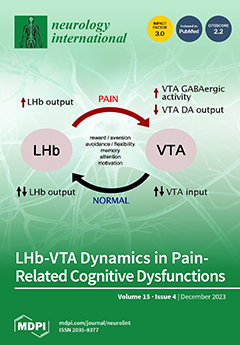
The coexistence of chronic pain and depressive symptoms forms an intrinsic relationship that affects executive and cognitive functions. This review delves into the pivotal role of the microcircuit formed between the lateral habenula (LHb) and the ventral tegmental area (VTA) in these dysfunctions. Dysregulation in VTA dopamine transmission, linked to LHb hyperactivity, may exacerbate cognitive issues related to pain. This review explores the organizational and structural connectivity aspects of the LHb-VTA microcircuit and its implications for pain-related cognitive dysfunction, offering potential avenues for targeted therapeutic interventions. View this paper
  • Issues are regarded as officially published after their release is announced to the table of contents alert mailing list.
  • You may sign up for e-mail alerts to receive table of contents of newly released issues.
  • PDF is the official format for papers published in both, html and pdf forms. To view the papers in pdf format, click on the "PDF Full-text" link, and use the free Adobe Reader to open them.
Order results
Result details
Select all
Export citation of selected articles as:
8 pages, 811 KiB  
Case Report
The Beneficial Outcome of Subsequent Treatment with Anakinra during the Chronic Phase of Febrile Infection-Related Epilepsy Syndrome (FIRES): A Case Report
Neurol. Int. 2023, 15(4), 1489-1496; https://doi.org/10.3390/neurolint15040097 - 11 Dec 2023
Viewed by 626
Abstract
This case report presents the clinical course of an eight-year-old boy diagnosed with febrile infection-related epilepsy syndrome (FIRES) at the age of four. Following a febrile infection, the patient experienced his initial episode of serial generalized clonic seizures. The severity of his condition [...] Read more.
This case report presents the clinical course of an eight-year-old boy diagnosed with febrile infection-related epilepsy syndrome (FIRES) at the age of four. Following a febrile infection, the patient experienced his initial episode of serial generalized clonic seizures. The severity of his condition led to 11 hospital admissions, totaling 157 days of hospitalization. Anakinra was initially administered during the acute phase in 2019 but was discontinued after 29 days. In 2022, the patient experienced a chronic-phase exacerbation and underwent a second course of anakinra treatment, which demonstrated a positive effect on seizure activity. With a year of anakinra therapy, the patient exhibited significant improvement in both seizure frequency and severity. This report adds to the existing evidence supporting the potential use of anakinra in the treatment of FIRES, highlighting its effectiveness during the chronic phase and suggesting the potential benefits of subsequent administration. Full article
Show Figures

Figure 1

9 pages, 288 KiB  
Article
Middle Meningeal Artery Embolization versus Surgery in Patients with Chronic Subdural Hematoma—No More Fence Sitting?
Neurol. Int. 2023, 15(4), 1480-1488; https://doi.org/10.3390/neurolint15040096 - 06 Dec 2023
Viewed by 481
Abstract
Background: Endovascular treatment of patients with chronic subdural hematoma using middle meningeal artery (MMA) embolization could become an alternative to surgical hematoma evacuation. The aim of the study was to compare methods and identify parameters to help determine the correct treatment modality. Methods: [...] Read more.
Background: Endovascular treatment of patients with chronic subdural hematoma using middle meningeal artery (MMA) embolization could become an alternative to surgical hematoma evacuation. The aim of the study was to compare methods and identify parameters to help determine the correct treatment modality. Methods: We retrospectively reviewed 142 cases conducted internally; 78 were treated surgically and 64 were treated using MMA embolization. We analyzed the treatment failure rate and complications, and using a binary logistic regression model, we identified treatment failure risk factors. Results: We found a comparable treatment failure rate of 23.1% for the surgery group and 21.9% for the MMA embolization group. However, in the MMA embolization group, 11 cases showed treatment failure due to early neurological worsening with a need for concomitant surgery. We also found a recurrence of hematoma in 15.4% of cases in the surgery group and 6.3% of cases in the MMA embolization group. Conclusion: Both modalities have their advantages; however, correct identification is crucial for treatment success. According to our findings, hematomas with a maximal width of <18 mm, a midline shift of <5 mm, and no acute or subacute (hyperdense) hematoma could be treated with MMA embolization. Hematomas with a maximal width of >18 mm, a midline shift of >5 mm, and no membranous segmentation could have better outcomes after surgical treatment. Full article
11 pages, 1899 KiB  
Article
Increased Susceptibility to Pilocarpine-Induced Status Epilepticus and Reduced Latency in TRPC1/4 Double Knockout Mice
Neurol. Int. 2023, 15(4), 1469-1479; https://doi.org/10.3390/neurolint15040095 - 06 Dec 2023
Viewed by 392
Abstract
Canonical transient receptor potential channels (TRPCs) are a family of calcium-permeable cation channels. Previous studies have shown that heteromeric channels comprising TRPC1 and TRPC4 mediate epileptiform bursting in lateral septal neurons and hippocampal CA1 pyramidal neurons, suggesting that TRPC1/4 channels play a pro-seizure [...] Read more.
Canonical transient receptor potential channels (TRPCs) are a family of calcium-permeable cation channels. Previous studies have shown that heteromeric channels comprising TRPC1 and TRPC4 mediate epileptiform bursting in lateral septal neurons and hippocampal CA1 pyramidal neurons, suggesting that TRPC1/4 channels play a pro-seizure role. In this study, we utilized electroencephalography (EEG) recording and spectral analysis to assess the role of TRPC1/4 channels in the pilocarpine model of status epilepticus (SE). We found that, surprisingly, TRPC1/4 double knockout (DKO) mice exhibited an increased susceptibility to pilocarpine-induced SE. Furthermore, SE latency was also significantly reduced in TRPC1/4 DKO mice. Further studies are needed to reveal the underlying mechanisms of our unexpected results. Full article
Show Figures

Figure 1

10 pages, 472 KiB  
Article
Physical Rehabilitation and Post-Stroke Pneumonia: A Retrospective Observational Study Using the Japanese Diagnosis Procedure Combination Database
Neurol. Int. 2023, 15(4), 1459-1468; https://doi.org/10.3390/neurolint15040094 - 04 Dec 2023
Viewed by 628
Abstract
In this study, the relationship between the duration of physical rehabilitation and occurrence of pneumonia after ischemic stroke was examined. We included 426,508 patients aged 75 years with acute ischemic stroke. A multilevel logistic regression analysis nested at the hospital level was [...] Read more.
In this study, the relationship between the duration of physical rehabilitation and occurrence of pneumonia after ischemic stroke was examined. We included 426,508 patients aged 75 years with acute ischemic stroke. A multilevel logistic regression analysis nested at the hospital level was conducted to examine the association between the duration of physical rehabilitation and occurrence of pneumonia. The duration of physical rehabilitation refers to the hours of physical rehabilitation performed daily until the 7th day of hospitalization. In the multivariable analysis, the intensity of rehabilitation for durations of 20–39 min/day (adjusted odds ratio [aOR]: 0.78, 95% Confidence Interval [CI]: 0.75–0.81, p < 0.001), 40–59 min/day (aOR: 0.68, 95% CI: 0.66–0.71, p < 0.001), 60–79 min/day (aOR:0.56, 95% CI: 0.53–0.58, p < 0.001), and 80 min/day (aOR: 0.46, 95% CI: 0.44–0.48, p < 0.001) were significantly associated with a reduced incidence of pneumonia. In addition, the trend identified for duration of rehabilitation was significant (p < 0.001). The results of this study suggest the usefulness of high-duration physical rehabilitation for preventing pneumonia in older patients with ischemic stroke. Full article
(This article belongs to the Special Issue Treatment Strategy and Mechanism of Acute Ischemic Stroke)
Show Figures

Graphical abstract

16 pages, 2052 KiB  
Review
Hemorrhagic Coagulation Disorders and Ischemic Stroke: How to Reconcile Both?
Neurol. Int. 2023, 15(4), 1443-1458; https://doi.org/10.3390/neurolint15040093 - 30 Nov 2023
Viewed by 331
Abstract
Coagulation and fibrinolytic system disorders are conditions in which the blood’s ability to clot is impaired, resulting in an increased risk of thrombosis or bleeding. Although these disorders are the expression of two opposing tendencies, they can often be associated with or be [...] Read more.
Coagulation and fibrinolytic system disorders are conditions in which the blood’s ability to clot is impaired, resulting in an increased risk of thrombosis or bleeding. Although these disorders are the expression of two opposing tendencies, they can often be associated with or be a consequence of each other, contributing to making the prognosis of acute cerebrovascular events more difficult. It is important to recognize those conditions that are characterized by dual alterations in the coagulation and fibrinolytic systems to reduce the prognostic impact of clinical conditions with difficult treatment and often unfortunate outcomes. Management of these individuals can be challenging, as clinicians must balance the need to prevent bleeding episodes with the potential risk of clot formation. Treatment decisions should be made on an individual basis, considering the specific bleeding disorder, its severity, and the patient’s general medical condition. This review aims to deal with all those forms in which coagulation and fibrinolysis represent two sides of the same media in the correct management of patients with acute neurological syndrome. Precision medicine, personalized treatment, advanced anticoagulant strategies, and innovations in bleeding control represent future directions in the management of these complex pathologies in which stroke can be the evolution of two different acute events or be the first manifestation of an occult or unknown underlying pathology. Full article
(This article belongs to the Special Issue Treatment Strategy and Mechanism of Acute Ischemic Stroke)
Show Figures

Figure 1

20 pages, 759 KiB  
Review
The Effects of Transcranial Direct Current Stimulation in Patients with Mild Cognitive Impairment
Neurol. Int. 2023, 15(4), 1423-1442; https://doi.org/10.3390/neurolint15040092 - 29 Nov 2023
Viewed by 576
Abstract
Transcranial direct current stimulation (tDCS) came into consideration in recent years as a promising, non-invasive form of neuromodulation for individuals suffering from mild cognitive impairment (MCI). MCI represents a transitional stage between normal cognitive aging and more severe cognitive decline, which appears in [...] Read more.
Transcranial direct current stimulation (tDCS) came into consideration in recent years as a promising, non-invasive form of neuromodulation for individuals suffering from mild cognitive impairment (MCI). MCI represents a transitional stage between normal cognitive aging and more severe cognitive decline, which appears in neurodegenerative diseases, such as Alzheimer’s disease. Numerous studies have shown that tDCS can have several useful effects in patients with MCI. It is believed to enhance cognitive functions, including memory and attention, potentially slowing down the progression of neurodegeneration and cognitive decline. tDCS is believed to work by modulating neuronal activity and promoting synaptic plasticity in the brain regions associated with cognition. Moreover, tDCS is generally considered safe and well-tolerated, making it an attractive option for long-term therapeutic use in MCI. However, further research is needed to determine the optimal stimulation parameters and long-term effects of tDCS in this population, as well as its potential to serve as a complementary therapy alongside other interventions for MCI. In this review, we included 16 randomized clinical trials containing patients with MCI who were treated with tDCS. We aim to provide important evidence for the cognitive enhancement using tDCS in patients with MCI, summarizing the effects and conclusions found in several clinical trials, and discuss its main mechanisms. Full article
Show Figures

Figure 1

12 pages, 1125 KiB  
Article
Video Motion Analysis as a Quantitative Evaluation Tool for Essential Tremor during Magnetic Resonance-Guided Focused Ultrasound Thalamotomy
Neurol. Int. 2023, 15(4), 1411-1422; https://doi.org/10.3390/neurolint15040091 - 29 Nov 2023
Viewed by 460
Abstract
The Clinical Rating Scale for Tremor (CRST) is commonly used to evaluate essential tremor (ET) during focused ultrasound (FUS) thalamotomy. However, it faces challenges such as the ceiling effect and test–retest variability. This study explored the utility of videographic motion analysis as an [...] Read more.
The Clinical Rating Scale for Tremor (CRST) is commonly used to evaluate essential tremor (ET) during focused ultrasound (FUS) thalamotomy. However, it faces challenges such as the ceiling effect and test–retest variability. This study explored the utility of videographic motion analysis as an evaluation index for ET. Forty-three patients with ET performed postural tremor and line-drawing tasks recorded on video, and the data were analyzed using motion analysis software. The test–retest and inter-rater reliability, correlations with the CRST and tremor scores, and pre/post-FUS treatment comparisons were analyzed. The video motion analysis showed excellent test–retest and inter-rater reliability. In the postural tremor tasks, video parameter amplitude significantly correlated with the CRST and tremor scores. Similarly, for the line-drawing task, video parameter amplitude showed significant correlations with CRST and tremor scores, effectively addressing the ceiling effect. Regarding post-FUS treatment improvements, changes in the CRST and tremor scores were significantly associated with changes in video parameter amplitude. In conclusion, quantitative analysis of the video motion of ET enables precise evaluation of kinematic characteristics and effectively resolves the ceiling effect and test–retest variability. The video motion analysis score accurately reflected the tremor severity and treatment effects, demonstrating its high clinical utility. Full article
Show Figures

Figure 1

8 pages, 2720 KiB  
Case Report
Astrocytoma Mimicking Herpetic Meningoencephalitis: The Role of Non-Invasive Multimodal Monitoring in Neurointensivism
Neurol. Int. 2023, 15(4), 1403-1410; https://doi.org/10.3390/neurolint15040090 - 29 Nov 2023
Viewed by 527
Abstract
Neuromonitoring is a critical tool for emergency rooms and intensive care units to promptly identify and treat brain injuries. The case report of a patient with status epilepticus necessitating orotracheal intubation and intravenous lorazepam administration is presented. A pattern of epileptiform activity was [...] Read more.
Neuromonitoring is a critical tool for emergency rooms and intensive care units to promptly identify and treat brain injuries. The case report of a patient with status epilepticus necessitating orotracheal intubation and intravenous lorazepam administration is presented. A pattern of epileptiform activity was detected in the left temporal region, and intravenous Acyclovir was administered based on the diagnostic hypothesis of herpetic meningoencephalitis. The neurointensivist opted for multimodal non-invasive bedside neuromonitoring due to the complexity of the patient’s condition. A Brain4care (B4C) non-invasive intracranial compliance monitor was utilized alongside the assessment of an optic nerve sheath diameter (ONSD) and transcranial Doppler (TCD). Based on the collected data, a diagnosis of intracranial hypertension (ICH) was made and a treatment plan was developed. After the neurosurgery team’s evaluation, a stereotaxic biopsy of the temporal lesion revealed a grade 2 diffuse astrocytoma, and an urgent total resection was performed. Research suggests that monitoring patients in a dedicated neurologic intensive care unit (Neuro ICU) can lead to improved outcomes and shorter hospital stays. In addition to being useful for patients with a primary brain injury, neuromonitoring may also be advantageous for those at risk of cerebral hemodynamic impairment. Lastly, it is essential to note that neuromonitoring technologies are non-invasive, less expensive, safe, and bedside-accessible approaches with significant diagnostic and monitoring potential for patients at risk of brain abnormalities. Multimodal neuromonitoring is a vital tool in critical care units for the identification and management of acute brain trauma as well as for patients at risk of cerebral hemodynamic impairment. Full article
Show Figures

Figure 1

10 pages, 306 KiB  
Article
A Characterization of Neurology Consults for Inpatients with SARS-CoV-2 Infection Compared to Other Respiratory Viruses
Neurol. Int. 2023, 15(4), 1393-1402; https://doi.org/10.3390/neurolint15040089 - 23 Nov 2023
Viewed by 503
Abstract
Introduction: Neurological consultation for patients infected with SARS-CoV-2 is common; it is currently unknown whether the neurologist’s approach to inpatient consultation of patients with SARS-CoV-2 should differ from the paradigm used to evaluate hospitalized patients with similar respiratory viruses. The goal of the [...] Read more.
Introduction: Neurological consultation for patients infected with SARS-CoV-2 is common; it is currently unknown whether the neurologist’s approach to inpatient consultation of patients with SARS-CoV-2 should differ from the paradigm used to evaluate hospitalized patients with similar respiratory viruses. The goal of the present study is to determine if the preponderance of new neurologic diagnoses differs between inpatients with SARS-CoV-2 and similar non-SARS-CoV-2 respiratory viruses for whom neurology is consulted. Methods: We performed a retrospective chart analysis of inpatient neurologic consultations at three major Philadelphia-based hospitals. We compared the final neurologic diagnosis of 152 patients infected with SARS-CoV-2 to 54 patients with a similar ubiquitous non-SARS-CoV-2 respiratory virus (influenza A, influenza B, respiratory syncytial virus, rhinovirus, or adenovirus, the most commonly tested respiratory viruses at our institution). Secondary metrics included age, sex, level of care, prior neurologic diagnoses, and mortality. A multinomial logistic regression model was utilized to evaluate the relative difference between diagnostic category groups on all metrics. Results: The proportion of patients with seizure who were infected with SARS-CoV-2 admitted to an intensive care unit (ICU) was significantly higher than those who were admitted to a medical–surgical floor. SARS-CoV-2 was also associated with increased risk for ICU admission compared to other common respiratory viruses. SARS-CoV-2 inpatients requiring neurologic consultation were also more likely to be older and female as compared to the non-SARS-CoV-2 cohort. In other domains, the proportion of neurologic diagnoses between SAR-CoV-2 and non-SARS-CoV-2 respiratory viruses showed no significant difference. Conclusion: Patients requiring inpatient neurologic consultation with a diagnosis of SARS-CoV-2 infection or another respiratory virus were found to be remarkably similar in terms of their ultimate neurologic diagnosis, with the exception of a larger preponderance of seizure in critical-care-level patients with SARS-CoV-2 infection. Our study suggests that the neurological approach to patients hospitalized with SARS-CoV-2 should be similar to that for patients with similar common respiratory infections, noting that seizure was seen more frequently in critically ill patients infected with SARS-CoV-2. Full article
(This article belongs to the Special Issue COVID-19, Neuroinflammation and Therapeutics)
10 pages, 2153 KiB  
Article
Characterizing the Impact of Compression Duration and Deformation-Related Loss of Closure Force on Clip-Induced Spinal Cord Injury in Rats
Neurol. Int. 2023, 15(4), 1383-1392; https://doi.org/10.3390/neurolint15040088 - 13 Nov 2023
Viewed by 523
Abstract
The clip-induced spinal cord injury (SCI) rat model is pivotal in preclinical SCI research. However, the literature exhibits variability in compression duration and limited attention to clip deformation-related loss of closure force. We aimed to investigate the impact of compression duration on SCI [...] Read more.
The clip-induced spinal cord injury (SCI) rat model is pivotal in preclinical SCI research. However, the literature exhibits variability in compression duration and limited attention to clip deformation-related loss of closure force. We aimed to investigate the impact of compression duration on SCI severity and the influence of clip deformation on closure force. Rats received T10-level clip-induced SCI with durations of 1, 5, 10, 20, and 30 s, and a separate group underwent T10 transection. Outcomes included functional, histological, electrophysiological assessments, and inflammatory cytokine analysis. A tactile pressure mapping system quantified clip closure force after open–close cycles. Our results showed a positive correlation between compression duration and the severity of functional, histological, and electrophysiological deficits. Remarkably, even a brief 1-s compression caused significant deficits comparable to moderate-to-severe SCI. SSEP waveforms were abolished with durations over 20 s. Decreased clip closure force appeared after five open–close cycles. This study offers critical insights into regulating SCI severity in rat models, aiding researchers. Understanding compression duration and clip fatigue is essential for experiment design and interpretation using the clip-induced SCI model. Full article
Show Figures

Figure 1

12 pages, 1252 KiB  
Article
Cognitive Failure in Adults with Spinal Cord Injury: A Valuable Adjunct Measure for Enhancing Cognitive Assessment and Rehabilitation Outcomes
Neurol. Int. 2023, 15(4), 1371-1382; https://doi.org/10.3390/neurolint15040087 - 08 Nov 2023
Viewed by 450
Abstract
Cognitive impairment is common in persons with spinal cord injury (SCI), impacting their daily functioning and rehabilitation. This study assesses the extent of self-reported cognitive failures in everyday life in persons with SCI and its relationships with objective neurocognitive measures and psychosocial factors, [...] Read more.
Cognitive impairment is common in persons with spinal cord injury (SCI), impacting their daily functioning and rehabilitation. This study assesses the extent of self-reported cognitive failures in everyday life in persons with SCI and its relationships with objective neurocognitive measures and psychosocial factors, including depressive mood, anxiety, perceived control, and fatigue. The differences between forty-one adults with a chronic SCI and forty-one able-bodied controls were examined. The participants completed the Cognitive Failures Questionnaire (CFQ) to assess cognitive failure and neurocognitive tests assessing attention and executive functions, as well as a psychosocial assessment. The SCI group reported higher cognitive failure rates than the able-bodied group (31.7% versus 19%, p > 0.05). Objective neurocognitive tests did not significantly correlate with the CFQ scores in either group. However, the CFQ scores were positively associated with most psychosocial factors, even after controlling for covariates. The CFQ scores were significantly associated with depressive mood in persons with SCI. These findings highlight the importance of incorporating self-reported cognitive measures into neurocognitive assessments and rehabilitation planning for adults with SCI. Self-reports capture everyday cognitive challenges that objective tests may miss. Additionally, this study highlights the strong connections between cognitive failures and psychosocial issues, particularly mood disorders, emphasizing the need for comprehensive rehabilitation and psychosocial support post-SCI, addressing both cognitive and emotional wellbeing. Full article
Show Figures

Figure 1

12 pages, 3663 KiB  
Article
Analysis of Post-COVID-19 Guillain–Barré Syndrome over a Period of One Year in the University Hospital of Split (Croatia)
Neurol. Int. 2023, 15(4), 1359-1370; https://doi.org/10.3390/neurolint15040086 - 06 Nov 2023
Viewed by 676
Abstract
The virus SARS-CoV-2 (Severe Acute Respiratory Syndrome Coronavirus 2) is capable of attacking the nervous system in several ways and leading to neurological diseases such as GBS (Guillain–Barré syndrome) through the resulting neurotropism and immune response. The aim of this study is to [...] Read more.
The virus SARS-CoV-2 (Severe Acute Respiratory Syndrome Coronavirus 2) is capable of attacking the nervous system in several ways and leading to neurological diseases such as GBS (Guillain–Barré syndrome) through the resulting neurotropism and immune response. The aim of this study is to show the relationship between Coronavirus disease (COVID-19) and GBS and to better understand the clinical symptoms to prevent poor outcomes. Data from 15 patients were extracted from the Department of Neurology, University Hospital of Split, Croatia, for the year 2021. The age of the patients ranged from 26 to 89 years, of whom 27% were women. Sixty seven percent of all GBS patients recovered from COVID-19 infection, whereas post-vaccinal polyradiculoneuritis was detected in 6%. Forty four percent of the patients who developed GBS had a severe form of COVID-19 infection. Forty percent of patients were treated with intravenous immunoglobulins (IVIG), followed by therapeutic plasma exchange (PLEX) in 27%. After the therapy, improvement was observed in 13 patients, while two patients died. The results suggest that SARS-CoV-2 triggers GBS because it follows a similar pattern of infection as the other viral and bacterial agents that contribute to the onset of GBS. There is no evidence that prior infection with COVID-19 worsens the clinical presentation of GBS. Full article
(This article belongs to the Special Issue Global Burden of Neurological Disorder)
Show Figures

Graphical abstract

7 pages, 1571 KiB  
Case Report
A Case Series of Four Patients with Artery of Percheron Occlusion over a Three-Month Period
Neurol. Int. 2023, 15(4), 1352-1358; https://doi.org/10.3390/neurolint15040085 - 02 Nov 2023
Viewed by 1052
Abstract
Here, we present a case series of four patients diagnosed with acute ischaemic stroke due to occlusion of the artery of Percheron (AOP), a rare stroke variant, observed in a single emergency centre within a three-month period. AOP occlusion is characterized by bilateral [...] Read more.
Here, we present a case series of four patients diagnosed with acute ischaemic stroke due to occlusion of the artery of Percheron (AOP), a rare stroke variant, observed in a single emergency centre within a three-month period. AOP occlusion is characterized by bilateral thalamic infarction with or without involvement of the mesencephalon. The presenting symptoms are diverse and not specific, but commonly include disturbance of consciousness, memory impairment, and vertical gaze palsy. In addition, due to the location of the infarction, imaging recognition is challenging and AOP occlusion often remains undiagnosed. This paper emphasizes the necessity of early recognition and appropriate management of AOP occlusion to significantly impact patient outcomes. Moreover, we argue that the condition might be more common than previously thought and that misdiagnosis or delay in diagnosis may lead to inappropriate treatment and potential failure to apply thrombolysis within the required timeframe. Full article
Show Figures

Figure 1

13 pages, 954 KiB  
Article
Fecal Incontinence after Severe Brain Injury: A Barrier to Discharge after Inpatient Rehabilitation?
Neurol. Int. 2023, 15(4), 1339-1351; https://doi.org/10.3390/neurolint15040084 - 31 Oct 2023
Viewed by 706
Abstract
Background: In this study, we aimed to investigate the incidence of fecal incontinence (FI) after severe acquired brain injuries (sABIs) and to determine whether this symptom can lead to an inability to return home after rehabilitation. Methods: This was a retrospective observational cohort [...] Read more.
Background: In this study, we aimed to investigate the incidence of fecal incontinence (FI) after severe acquired brain injuries (sABIs) and to determine whether this symptom can lead to an inability to return home after rehabilitation. Methods: This was a retrospective observational cohort study. In total, 521 acute sABI inpatients were enrolled from the Department of Neurorehabilitation at an academic tertiary care hospital. Patients were divided into two groups, with and without FI, at the end of the rehabilitation phase. The primary and secondary endpoints were the incidence of persistent FI and any difference in the discharge destination. Results: Upon admission, new-onset FI was found in 443 (85%) patients, of which 38% had traumatic sABI. Moreover, 62.7% of all patients had FI upon admission. At discharge, 53.3% (264/495) of patients still had FI. Of these, 75.4% (199/264) had a Rancho Level of Cognitive Functioning Scale (LCFS) ≥3. A statistically significant correlation between FI at discharge and the presence of frontal lesions, autonomic crises, and increased LCFS scores was noted. Among the patients discharged to their homes, the proportion with persistent FI was lower (34% vs. 53.3). Conclusions: FI was significantly persistent after sABI, even after recovery from unconsciousness, and must be considered as a consequence of, rather than an independent risk factor for, unfavorable outcomes. Full article
(This article belongs to the Special Issue Global Burden of Neurological Disorder)
Show Figures

Figure 1

19 pages, 2413 KiB  
Article
Remdesivir-Loaded Nanoliposomes Stabilized by Chitosan/Hyaluronic Acid Film with a Potential Application in the Treatment of Coronavirus Infection
Neurol. Int. 2023, 15(4), 1320-1338; https://doi.org/10.3390/neurolint15040083 - 30 Oct 2023
Viewed by 621
Abstract
An object of the present study was the development of liposomes loaded with the medicine Veklury® (remdesivir) stabilized by electrostatic adsorption of polysaccharide film formed from chitosans with different physicochemical characteristics and hyaluronic acid. The functionalization of the structures was achieved through [...] Read more.
An object of the present study was the development of liposomes loaded with the medicine Veklury® (remdesivir) stabilized by electrostatic adsorption of polysaccharide film formed from chitosans with different physicochemical characteristics and hyaluronic acid. The functionalization of the structures was achieved through the inclusion of an aptamer (oligonucleotide sequence) with specific affinity to the spike protein of the human coronavirus HCoV-OC43. The hydrodynamic size, electrokinetic potential and stability of the structures were evaluated at each step in the procedure. The encapsulation efficiency and loaded amount of remdesivir (99% and 299 µg/mL) were estimated by UV–vis spectroscopy. Our investigations showed manifestation of promising tendencies for prolonged periods of the drug release and increased effectiveness of its antiviral action. Among all studied versions of the delivery system, the most distinguished and suitable in a model coronavirus therapy are the liposomes formed from chitosan oligosaccharides. The cytotoxicity of the liposomes was determined against the HCT-8 cell line. A cytopathic effect inhibition test was used for the assessment of the antiviral activity of the compounds. The virucidal activity and the effect on the viral adsorption of the samples were reported by the end-point dilution method, and the alteration in viral titer was determined as Δlgs compared to untreated controls. The redox-modulating properties of the nanoparticles were studied in vitro in certain/several/a few chemical model systems. Our investigations showed a manifestation of promising tendencies for a prolonged effect of the drug release and increased effectiveness of its antiviral action. Full article
(This article belongs to the Special Issue COVID-19, Neuroinflammation and Therapeutics)
Show Figures

Figure 1

17 pages, 705 KiB  
Review
Dynamics of Lateral Habenula–Ventral Tegmental Area Microcircuit on Pain-Related Cognitive Dysfunctions
Neurol. Int. 2023, 15(4), 1303-1319; https://doi.org/10.3390/neurolint15040082 - 27 Oct 2023
Viewed by 756
Abstract
Chronic pain is a health problem that affects the ability to work and perform other activities, and it generally worsens over time. Understanding the complex pain interaction with brain circuits could help predict which patients are at risk of developing central dysfunctions. Increasing [...] Read more.
Chronic pain is a health problem that affects the ability to work and perform other activities, and it generally worsens over time. Understanding the complex pain interaction with brain circuits could help predict which patients are at risk of developing central dysfunctions. Increasing evidence from preclinical and clinical studies suggests that aberrant activity of the lateral habenula (LHb) is associated with depressive symptoms characterized by excessive negative focus, leading to high-level cognitive dysfunctions. The primary output region of the LHb is the ventral tegmental area (VTA), through a bidirectional connection. Recently, there has been growing interest in the complex interactions between the LHb and VTA, particularly regarding their crucial roles in behavior regulation and their potential involvement in the pathological impact of chronic pain on cognitive functions. In this review, we briefly discuss the structural and functional roles of the LHb–VTA microcircuit and their impact on cognition and mood disorders in order to support future studies addressing brain plasticity during chronic pain conditions. Full article
Show Figures

Graphical abstract

13 pages, 1281 KiB  
Article
Effects of Visual–Motor Illusion via Image Videos Showing Increased Exercise Intensity on the Tibial Anterior during Sit-to-Stand Movement: A Study of Healthy Participants
Neurol. Int. 2023, 15(4), 1290-1302; https://doi.org/10.3390/neurolint15040081 - 20 Oct 2023
Viewed by 635
Abstract
Visual–motor illusion (VMI) elicits kinesthetic sensation from visual stimulation. We have previously performed ankle motion VMI with resistance applied to the ankle joint on the paralyzed side (power-VMI (P-VMI)) and ankle motion VMI without resistance (standard-VMI (S-VMI)) to activate the tibialis anterior (TA) [...] Read more.
Visual–motor illusion (VMI) elicits kinesthetic sensation from visual stimulation. We have previously performed ankle motion VMI with resistance applied to the ankle joint on the paralyzed side (power-VMI (P-VMI)) and ankle motion VMI without resistance (standard-VMI (S-VMI)) to activate the tibialis anterior (TA) muscle in stroke-paralyzed patients and compared sit-to-stand (STS) durations, but these studies did not measure TA activity during the STS movement. The purpose of this study was to evaluate the effects of different intensities of visual stimuli presented during VMI on TA and STS movement. Healthy right-footed adults (n = 18) observed two different VMI videos of ankle dorsiflexion, including S-VMI and P-VMI, with an observation time of 2 min each. STS movement was evaluated before and after watching each video. Each participant performed both S-VMI and P-VMI interventions on the same day. Only P-VMI enhanced the integrated electromyogram of the TA, increased the angular velocities of the trunk forward inclination and the ankle dorsiflexion, and shortened the STS duration. Our results indicate that P-VMI facilitates the activation of TA during STS, and we believe that we have clarified the intervention mechanism of VMI. Full article
Show Figures

Figure 1

17 pages, 11685 KiB  
Case Report
Acute Presentation and Long-Term Rehabilitation Follow-Up of Ischemic Myelopathy Due to Clinically Suspected Fibrocartilaginous Embolism in an Adolescent Male: A Case Report and Review
Neurol. Int. 2023, 15(4), 1273-1289; https://doi.org/10.3390/neurolint15040080 - 19 Oct 2023
Viewed by 799
Abstract
Ischemic myelopathy is uncommon in the pediatric population, with fibrocartilaginous embolism (FCE) being one of its rarest causes. We present the case of an otherwise healthy 17-year-old student who experienced sudden onset of severe low-back pain amidst intensive physical training, which rapidly deteriorated [...] Read more.
Ischemic myelopathy is uncommon in the pediatric population, with fibrocartilaginous embolism (FCE) being one of its rarest causes. We present the case of an otherwise healthy 17-year-old student who experienced sudden onset of severe low-back pain amidst intensive physical training, which rapidly deteriorated to complete sensory-motor paralysis of his lower limbs. He was treated with IV Methylprednisolone and anticoagulation after the initial work-up suggested spinal cord infarction. After eight days, sufficient clinical-radiological correlation was achieved to support FCE diagnosis as the most likely cause of infarction. He subsequently received inpatient rehabilitation treatment for four months, after which he was followed as an outpatient for a total period of 16 months. While significant neurological and functional gains were achieved during this period, he also experienced some worsening. This case highlights the importance both of performing a thorough assessment and being familiar with FCE as a possible differential diagnosis of spinal cord infarction in children, to facilitate its timely identification and proper acute and long-term management. This case report was prepared following CARE guidelines after obtaining the patient’s written informed consent. Full article
Show Figures

Figure 1

20 pages, 1794 KiB  
Review
Physiological Mechanisms Inherent to Diabetes Involved in the Development of Dementia: Alzheimer’s Disease
Neurol. Int. 2023, 15(4), 1253-1272; https://doi.org/10.3390/neurolint15040079 - 10 Oct 2023
Viewed by 875
Abstract
Type 2 diabetes mellitus (T2D) is a metabolic disease reaching pandemic levels worldwide. In parallel, Alzheimer’s disease (AD) and vascular dementia (VaD) are the two leading causes of dementia in an increasingly long-living Western society. Numerous epidemiological studies support the role of T2D [...] Read more.
Type 2 diabetes mellitus (T2D) is a metabolic disease reaching pandemic levels worldwide. In parallel, Alzheimer’s disease (AD) and vascular dementia (VaD) are the two leading causes of dementia in an increasingly long-living Western society. Numerous epidemiological studies support the role of T2D as a risk factor for the development of dementia. However, few basic science studies have focused on the possible mechanisms involved in this relationship. On the other hand, this review of the literature also aims to explore the relationship between T2D, AD and VaD. The data found show that there are several alterations in the central nervous system that may be promoting the development of T2D. In addition, there are some mechanisms by which T2D may contribute to the development of neurodegenerative diseases such as AD or VaD. Full article
(This article belongs to the Topic Translational Advances in Neurodegenerative Dementias)
Show Figures

Figure 1

15 pages, 727 KiB  
Article
Headache and NOTCH3 Gene Variants in Patients with CADASIL
Neurol. Int. 2023, 15(4), 1238-1252; https://doi.org/10.3390/neurolint15040078 - 09 Oct 2023
Viewed by 1149
Abstract
Autosomal dominant cerebral arteriopathy with subcortical infarcts and leukoencephalopathy (CADASIL) is an inherited vascular disease characterized by recurrent strokes, cognitive impairment, psychiatric symptoms, apathy, and migraine. Approximately 40% of patients with CADASIL experience migraine with aura (MA). In addition to MA, CADASIL patients [...] Read more.
Autosomal dominant cerebral arteriopathy with subcortical infarcts and leukoencephalopathy (CADASIL) is an inherited vascular disease characterized by recurrent strokes, cognitive impairment, psychiatric symptoms, apathy, and migraine. Approximately 40% of patients with CADASIL experience migraine with aura (MA). In addition to MA, CADASIL patients are described in the literature as having migraine without aura (MO) and other types of headaches. Mutations in the NOTCH3 gene cause CADASIL. This study investigated NOTCH3 genetic variants in CADASIL patients and their potential association with headache types. Genetic tests were performed on 30 patients with CADASIL (20 women aged 43.6 ± 11.5 and 10 men aged 39.6 ± 15.8). PCR-HRM and sequencing methods were used in the genetic study. We described three variants as pathogenic/likely pathogenic (p.Tyr189Cys, p.Arg153Cys, p.Cys144Arg) and two benign variants (p.Ala202=, p.Thr101=) in the NOTCH3 gene and also presented the NOTCH3 gene variant (chr19:15192258 G>T), which has not been previously described in the literature. Patients with pathogenic/likely pathogenic variants had similar headache courses. People with benign variants showed a more diverse clinical picture. It seems that different NOTCH3 variants may contribute to the differential presentation of a CADASIL headache, highlighting the diagnostic and prognostic value of headache characteristics in this disease. Full article
Show Figures

Graphical abstract

11 pages, 425 KiB  
Article
Deterioration of Cough, Respiratory, and Vocal Cord Functions in Patients with Multiple System Atrophy
Neurol. Int. 2023, 15(4), 1227-1237; https://doi.org/10.3390/neurolint15040077 - 02 Oct 2023
Viewed by 983
Abstract
The purpose of this study was to clarify changes in cough function in patients with multiple system atrophy (MSA). Seventeen probable patients with MSA were studied. Peak cough flow (PCF), respiratory function (percentage of vital capacity, percentage of forced vital capacity, and percentage [...] Read more.
The purpose of this study was to clarify changes in cough function in patients with multiple system atrophy (MSA). Seventeen probable patients with MSA were studied. Peak cough flow (PCF), respiratory function (percentage of vital capacity, percentage of forced vital capacity, and percentage of predicted forced expiratory volume in one second), respiratory muscle strength (percentage of maximal inspiratory mouth pressure and percentage of maximal expiratory mouth pressure), and maximum phonation time (MPT) were assessed. Walking ability, disease duration, possibility of air stacking, Unified MSA Rating Scale (UMSARS), and Movement Disorder Society Unified Parkinson’s Disease Rating Scale (MDS-UPDRS) Part III were also assessed. Data were separately analyzed for ambulatory and non-ambulatory groups categorized by Functional Ambulation Categories. PCF, respiratory function, respiratory muscle strength, and MPT were significantly lower in the non-ambulatory group than in the ambulatory group. On the other hand, no correlation between PCF and disease duration was observed. A significant number of patients in the non-ambulatory group were unable to hold their breath. The UMSARS and MDS-UPDRS Part III in the non-ambulatory group were significantly higher than in the ambulatory group. It was concluded that ambulatory dysfunction is associated with the decline of cough function and respiratory-related function in patients with MSA. Full article
Show Figures

Figure 1

15 pages, 861 KiB  
Review
Targeting Neutrophil Extracellular Traps for Stroke Prognosis: A Promising Path
Neurol. Int. 2023, 15(4), 1212-1226; https://doi.org/10.3390/neurolint15040076 - 28 Sep 2023
Viewed by 682
Abstract
Stroke has become the first cause of functional disability and one of the leading causes of mortality worldwide. Therefore, it is of crucial importance to develop accurate biomarkers to assess stroke risk and prognosis. Emerging evidence suggests that neutrophil extracellular trap (NET) levels [...] Read more.
Stroke has become the first cause of functional disability and one of the leading causes of mortality worldwide. Therefore, it is of crucial importance to develop accurate biomarkers to assess stroke risk and prognosis. Emerging evidence suggests that neutrophil extracellular trap (NET) levels may serve as a valuable biomarker to predict stroke occurrence and functional outcome. NETs are known to create a procoagulant state by serving as a scaffold for tissue factor (TF) and platelets inducing thrombosis by activating coagulation pathways and endothelium. A literature search was conducted in two databases (MEDLINE and Scopus) to trace all relevant studies published between 1 January 2016 and 31 December 2022, addressing the potential utility of NETs as a stroke biomarker. Only full-text articles in English were included. The current review includes thirty-three papers. Elevated NET levels in plasma and thrombi seem to be associated with increased mortality and worse functional outcomes in stroke, with all acute ischemic stroke, intracerebral hemorrhage, and subarachnoid hemorrhage included. Additionally, higher NET levels seem to correlate with worse outcomes after recanalization therapies and are more frequently found in strokes of cardioembolic or cryptogenic origin. Additionally, total neutrophil count in plasma seems also to correlate with stroke severity. Overall, NETs may be a promising predictive tool to assess stroke severity, functional outcome, and response to recanalization therapies. Full article
(This article belongs to the Collection Advances in Neurodegenerative Diseases)
Show Figures

Figure 1

12 pages, 1201 KiB  
Systematic Review
An Ecchordosis Physaliphora, a Rare Entity, Involving the Central Nervous System: A Systematic Review of the Literature
Neurol. Int. 2023, 15(4), 1200-1211; https://doi.org/10.3390/neurolint15040075 - 26 Sep 2023
Viewed by 1157
Abstract
Ecchordosis physaliphora (EP) is a benign notochordal remnant, which is often an incidental finding; however, it can rarely present with neurological symptoms. We performed a systematic review of the literature for cases of symptomatic EP published in PubMed, Web of Science and Embase [...] Read more.
Ecchordosis physaliphora (EP) is a benign notochordal remnant, which is often an incidental finding; however, it can rarely present with neurological symptoms. We performed a systematic review of the literature for cases of symptomatic EP published in PubMed, Web of Science and Embase from January 1982 to May 2023. This is the largest review to date and revealed 60 cases including ours. Headache (55%) and CSF rhinorrhea (32%) were the most frequent clinical manifestations. The majority of symptomatic EP lesions were located in the prepontine region (77%) and required surgical resection (75%). EP should be considered in patients with neurologic symptoms in the setting of prepontine or posterior sphenoid sinus lesions. While symptomatic patients often require surgical intervention, rare cases may respond to oral corticosteroids. Full article
Show Figures

Figure 1

Previous Issue
Next Issue
Back to TopTop