Article

Three New Species of Russulaceae (Russulales, Basidiomycota) from Southern China

1
State Key Laboratory of Ecological Pest Control for Fujian and Taiwan Crops, College of Life Sciences, Fujian Agriculture and Forestry University, Fuzhou 350002, China
2
Department of Biological Sciences, University of Illinois, Chicago, IL 60607, USA
3
College of Horticulture, Fujian Agriculture and Forestry University, Fuzhou 350002, China
4
Bureau of Fujian Junzifeng National Nature Reserve, Sanming 365200, China
5
Institute of Crop Sciences, Fujian Academy of Agricultural Sciences, Fuzhou 350013, China
*
Authors to whom correspondence should be addressed.
J. Fungi 2024, 10(1), 70; https://doi.org/10.3390/jof10010070 (registering DOI)
Submission received: 5 December 2023 / Revised: 27 December 2023 / Accepted: 11 January 2024 / Published: 15 January 2024

Abstract

:
The characterization of natural fungal diversity impacts our understanding of ecological and evolutionary processes and can lead to novel bioproduct discovery. Russula and Lactarius, both in the order Russulales, represent two large genera of ectomycorrhizal fungi that include edible as well as toxic varieties. Based on morphological and phylogenetic analyses, including nucleotide sequences of the internal transcribed spacer (ITS), the 28S large subunit of ribosomal RNA (LSU), the second largest subunit of RNA polymerase II (RPB2), the ribosomal mitochondrial small subunit (mtSSU), and the translation elongation factor 1-α (TEF1-α) gene sequences, we here describe and illustrate two new species of Russula and one new species of Lactarius from southern China. These three new species are: R. junzifengensis (R. subsect. Virescentinae), R. zonatus (R. subsect. Crassotunicatae), and L. jianyangensis (L. subsect. Zonarii).

1. Introduction

Fungi classified within the order Russulaceae include members of some of the most significant ectomycorrhizal genera found in almost all forest ecosystems, spanning across temperate, subtropical, and tropical regions [1]. The proliferation and ecological relevance of Russulaceae are evident in the lush forests of southern China and various other regions dispersed throughout South Asia. This fungal assemblage plays a pivotal role in mycorrhizal associations, contributing significantly to the vitality and sustainability of forest ecosystems [2,3]. Russulaceae establish intimate associations with various host plants [4,5]. In addition, these fungi have significant medicinal, nutritional, and bioremediation value, including as resources for novel drug discovery [6,7,8]. Fujian, a coastal province in southern China, is surrounded by mountains on three sides and the sea on the other. The subtropical monsoon climate in this region results in relatively warm, short winters and long, rainy summers compared to northern China [9,10]. Botanically, Fujian is positioned at the southernmost end of the Sino-Japanese Floristic Region and faces Taiwan across the sea, the latter of which belongs to the Indo-Malay Region [11]. The main mountain ranges in Fujian include the Wuyi, Shanling, Jiufeng, and Tailao (elevation of ~200–2158 m), which house subtropical evergreen broadleaf forests, mixed coniferous and broadleaf forests, and South Asian tropical rainforests. The major tree species in Fujian comprise Masson’s pine, bamboo groves, willow trees, banyans, and camphor [12], among which the Chinese fir, Chinese yew, Fujian pine, and Chinese swamp cypress are indigenous to Fujian. The diverse and unique local tree species found in Fujian, coupled with the warm and humid climate, are likely important factors conducive to the proliferation of their associated Russulaceae fungi.
The genus Russula Pers. (Russulaceae, Russulales, Basidiomycota) was established by Persoon in 1796 [13]. Members of this genus often constitute crucial components of forest ecosystems via their extensive associations with plants, and also likely as a food source (their fruiting bodies/mushrooms) for a variety of animals [14]. Indeed, a number of Russula species are globally recognized as edible fungi [15] and have displayed promising (biopharmaceutical) properties with respect to possessing anticancer and antioxidative activities [16,17]. The morphological classification system for Russula is characterized by brightly colored fragile caps, brittle context, amyloid warty spores, abundant sphaerocysts in a heteromerous trama, an absence of latex, and simple-septate hyphae [18,19]. The documented number of species cataloged within the genus Russula currently surpasses > 2000, with their fruiting bodies encompassing a vast array of variations in color, morphology, and anatomical characteristics. However, due to the substantial variability exhibited within this taxonomic group, many species still pose considerable challenges in terms of their accurate identification and differentiation. This complexity underscores the likelihood of new species awaiting detection and classification through attempts to compare molecular phylogenetic reconstruction with modern infrageneric classification [20]. Consequently, challenges persist in differentiating and taxonomically categorizing Russula species within fungal surveys and ethnopharmacological investigations [21].
The genus Lactarius also belongs to the family Russulaceae in the order Russulales [22]. In traditional classification, all species that exude latex (or “milk”, hence the term “milkcap” fungi) were grouped under the genus Lactarius. Buyck et al. [22,23] separated Lactifluus and L. furcatus Coker from the genus Lactarius, establishing a new genus named Lactarius sensu novo, which is mainly classified into three subgenera: L. subg. Lactarius, L. subg. Plinthogalus (Burl.) Hesler & A.H. Sm., and L. subg. Russularia (Fr.) Kauffman. Although the exploration of extensive fungal resources has led to the identification of several dozen Russulaceae species across various regions [24,25,26,27], investigations concerning this genus in southern China remain inadequately addressed, with the continual discovery of new species [28,29].
During an exploration aimed at delineating the diversity and geographical distribution of Russula in China, a series of intriguing samples was gathered within Fujian province, China. These isolates displayed characteristics that did not correspond to any known species within the genus. Employing both morphological and molecular phylogenetic analyses, we identify three new species within the Russulaceae family. We present detailed descriptions of these newfound species, complemented by illustrations elucidating their distinctive morphological attributes.

2. Materials and Methods

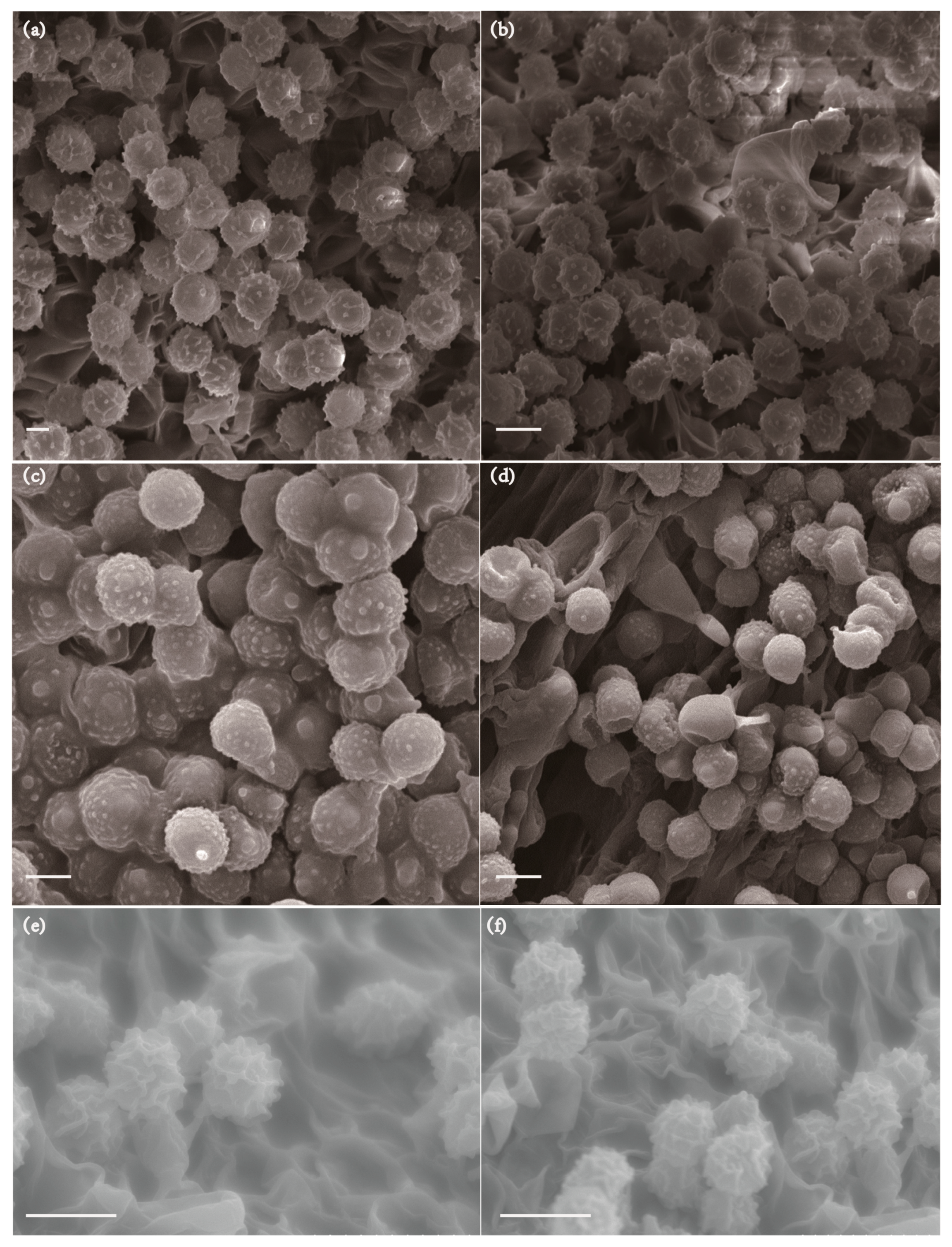
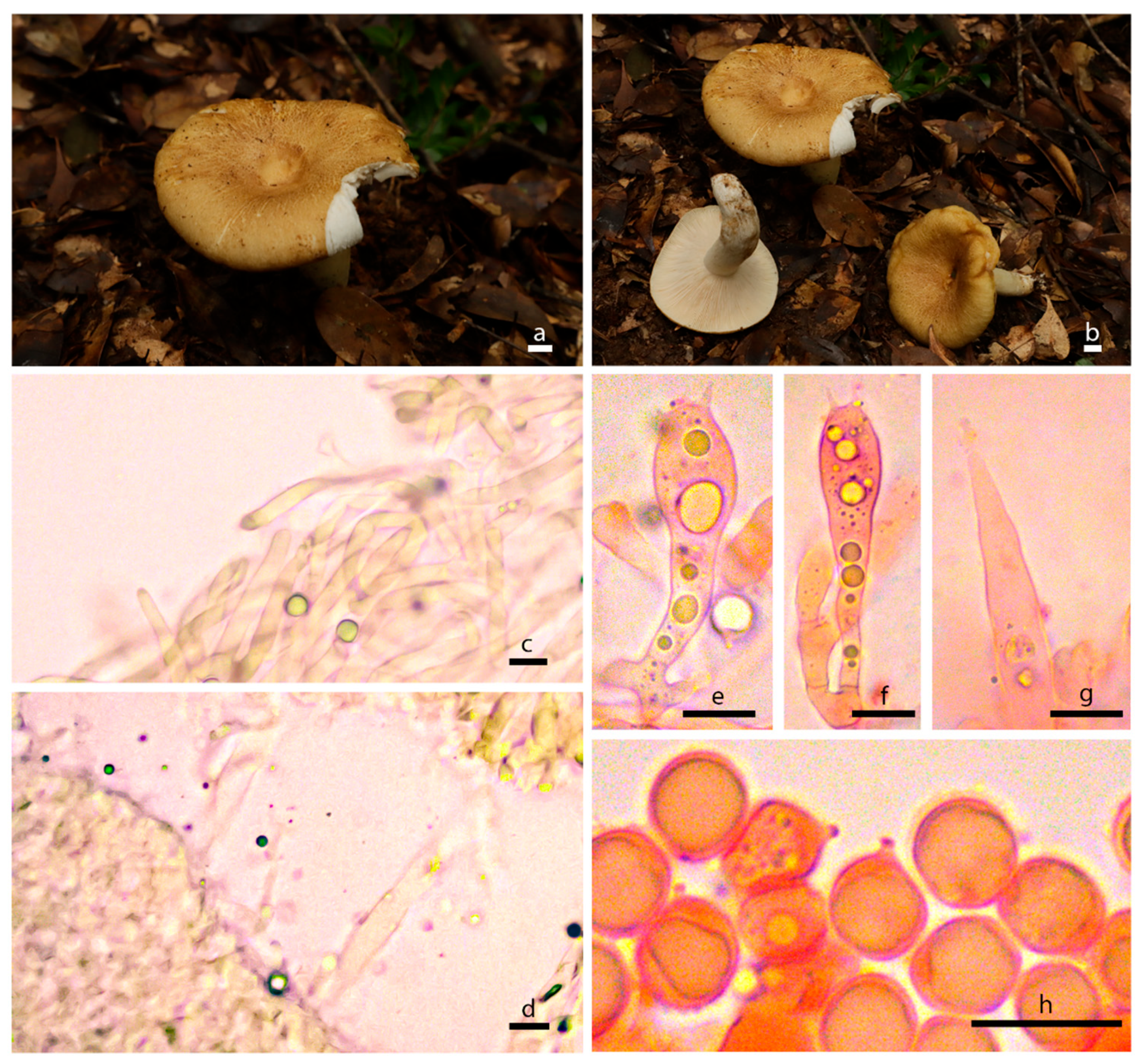
2.1. Collections and Morphological Analyses

Fresh fruiting bodies of two unknown (putative members of the Russula) mushrooms were collected from the Junzifeng National Nature Reserve, and one from Jiufeng Mountain, Jianyang (putative member of the Lactarius), in the Fujian Province, China, in August 2021. These specimens were collected during field expeditions focused on fungi. Images of the fresh fruiting bodies were captured using a Canon (Tokyo, Japan) EOS 6D Mark II camera. The meticulous documentation of their macroscopic attributes involved the careful examination of fresh samples in their natural diurnal environment. Comprehensive records encompassing macroscopic characteristics and habitat specifics were meticulously collated from collection records and accompanying visual documentation, adhering to the conventions of mycological taxonomic research. To ensure the permanent preservation of specimens, one crucial step was a dehydration process, during which the specimens underwent desiccation within a drying oven set at 45 °C. This meticulous procedure persisted until the moisture content of the fruiting bodies was diligently reduced to below 10%, ensuring their suitability for long-term storage. Microsections of dried specimens were stained with a mixture of 5% potassium hydroxide (KOH) and 1% Congo red. A detailed illustration of the structure and ornamentation of the spores was carried out using a scanning electron microscope (ZEM15C, ZEPTOOLS, Tongling, China). Microscopic features were observed using a Leica microscope (DM2500, Wetzlar, Germany) at magnifications up to 100×. For the description of basidiospores, 20 basidiospores, in profile view, were measured. The basidium length excludes sterigmata. The notation (a-)b–c(-d) was used to represent the dimensions of basidiospores, where the range ‘b–c’ covered 90% or more of the measured values. ‘a’ and ‘d’ represent the extreme values. An average length/width ratio (Q value) was calculated from 20 spores, along with the standard deviation, reflecting the characteristics of the basidiospores. The specimens were deposited in the Herbarium Mycologicum Academiae Sinicae (HMAS) at the Institute of Microbiology, Chinese Academy of Sciences, with the specimen numbers HMAS 298099, HMAS 298100, HMAS 298101, HMAS 298102, HMAS 298103, and HMAS 298104. Taxonomic information on the new taxa was submitted to MycoBank (http://www.mycobank.org (accessed on 10 January 2024)).

2.2. DNA Extraction, PCR Amplification, and Sequencing

DNA was extracted using the Fungal DNA Mini Kit (OMEGA-D3390, Feiyang Biological Engineering Corporation, Guangzhou, China) following the manufacturer’s protocol. Briefly, 100 mg of starting material (fruiting body) was used for DNA extraction. The amplification of the nucleotide sequences of the internal transcribed spacer (ITS), 28S large subunit regions of ribosomal DNA (LSU), the translation elongation factor 1-α (TEF1-α), the ribosomal mitochondrial small subunit (mtSSU), and second largest RNA polymerase II regions (RPB2) was conducted via polymerase chain reaction (PCR) using the primer pairs: ITS4/ITS5 [30], LROR/LR5 [31], TEF1-α [32], mtSSU [30], and RPB2-6F/RPB2-7cR [33], respectively. The PCR reaction volume was 25 μL, comprising 12.5 μL of 2× Rapid Taq Master Mix (Vazyme, Nanjing, China), 1 μL of each forward and reverse primer (10 μM) (Sangon, Shanghai, China), and 1 μL of template genomic DNA. The reaction mixture was adjusted to a total volume of 25 μL using distilled deionized water. Amplification products were visualized using 1% agarose gel electrophoresis. Sequencing was performed by Fuzhou Tsingke Company (Fuzhou, China) using bidirectional (double-stranded) sequencing.

2.3. Alignment and Phylogenetic Analyses

To construct the phylogenetic tree of Russulaceae, we utilized sequences obtained from six fungal strains and reference sequences for multi-locus phylogenetic analyses which were obtained from Rehner and Buckley [32], Chen et al. [34], Deng et al. [35], Buyck et al. [36], and Roy et al. [37]. The newly generated sequences were screened for similarity through a GenBank BLAST search. The ITS, LSU, mtSSU, RPB2, and TEF1-α sequences were aligned using the MAFFT v. 7.11 online tool (https://mafft.cbrc.jp/alignment/software/ (accessed on 23 December 2023)), followed by manual adjustments in MEGA 7.0. Phylogenetic analyses employed both maximum likelihood (ML) and Bayesian inference (BI) methods. ML analysis was conducted using RaxML-HPC2 on XSEDE v. 8.2.12 via the CIPRES Science Gateway portal, while BI analysis was performed using MrBayes on XSEDE v. 3.2.7a (https://www.phylo.org/ (accessed on 25 December 2023)). The consensus tree was constructed using FigTree v. 1.4.4 and further refined using Adobe Illustrator CS 6.0. Newly generated sequences from this study have been deposited in GenBank. Branches showing ML bootstrap support values (≥70) and Bayesian posterior probability (≥0.90) were considered significantly supported.

3. Results

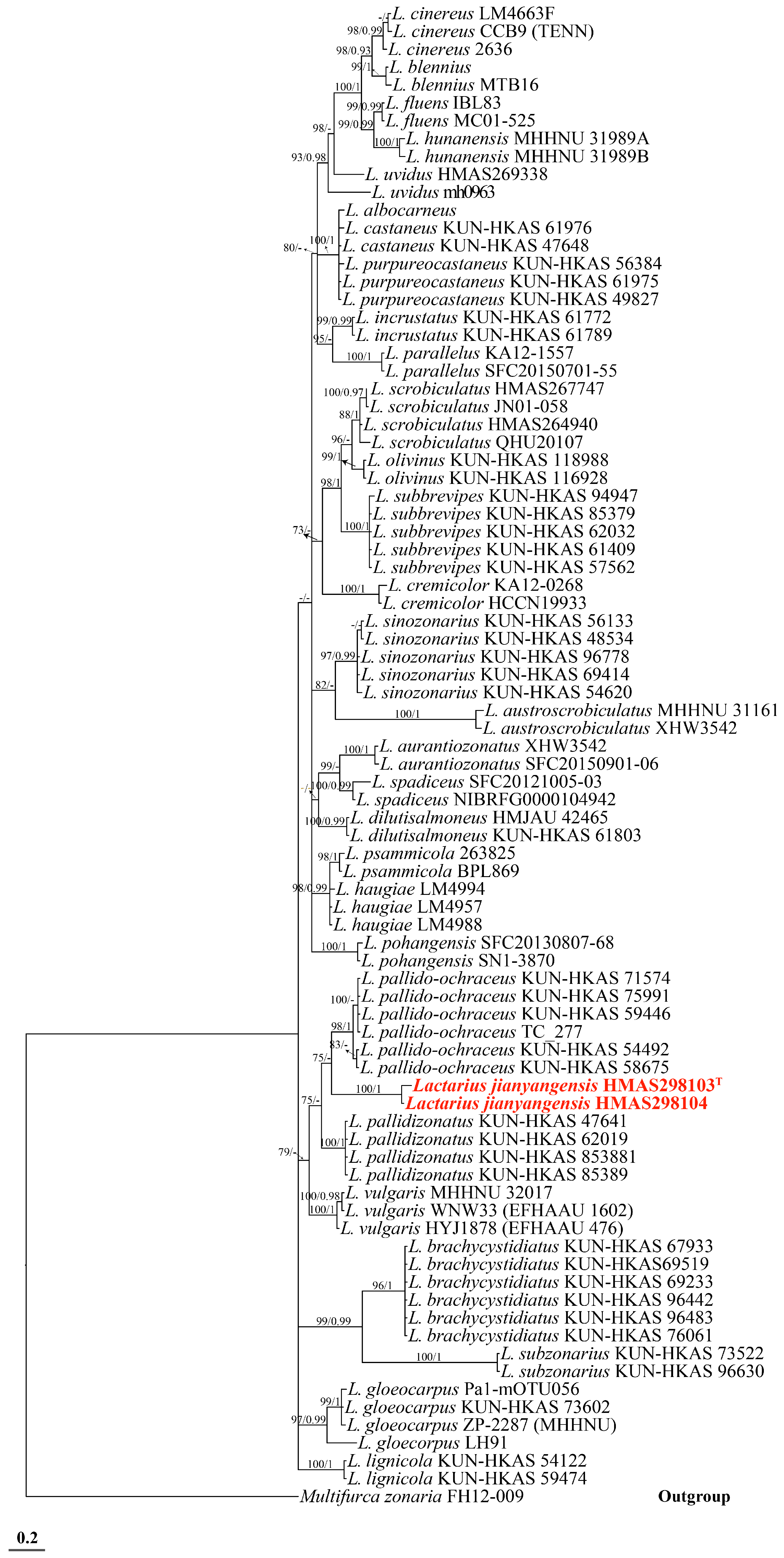
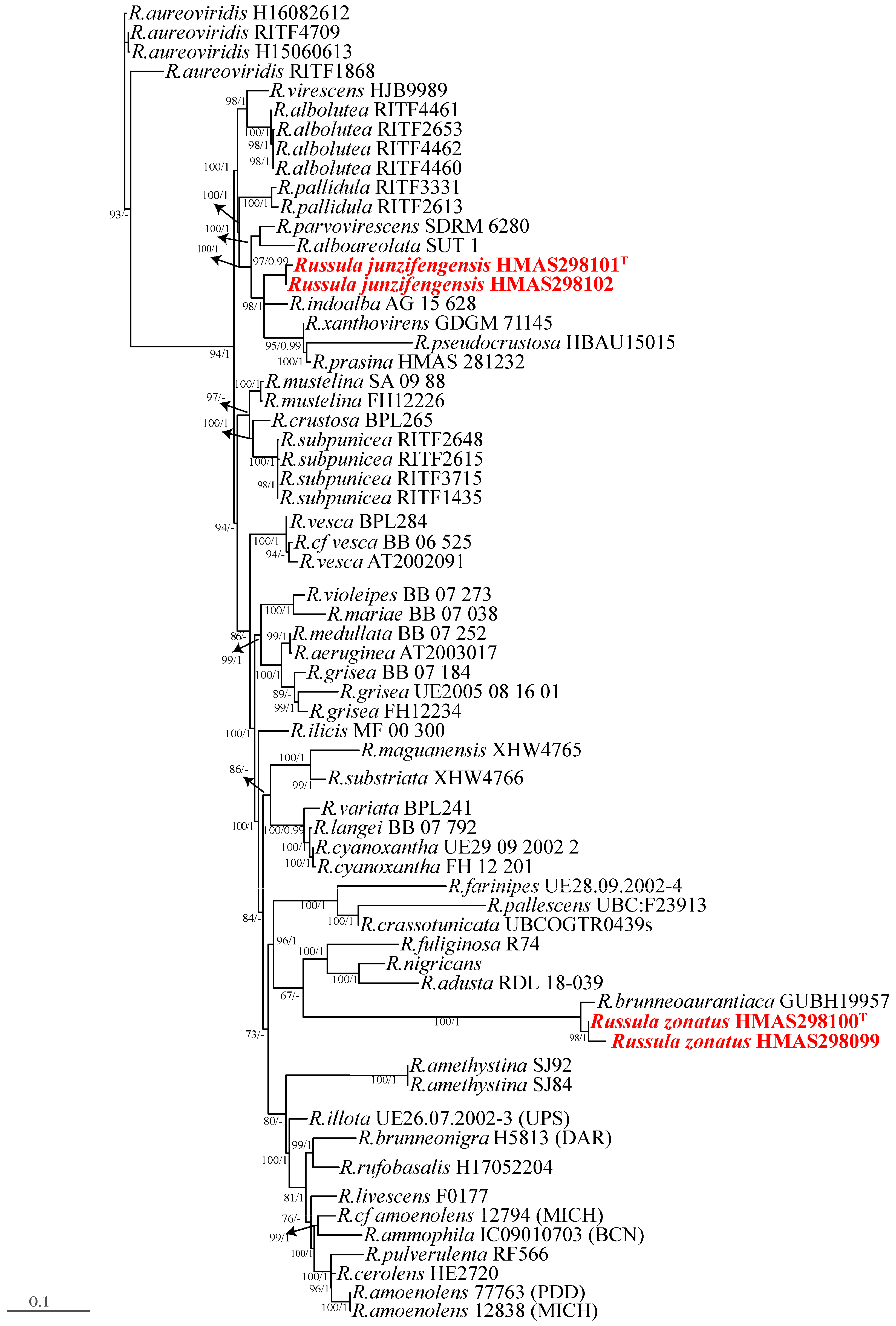
3.1. Phylogenetic Analyses
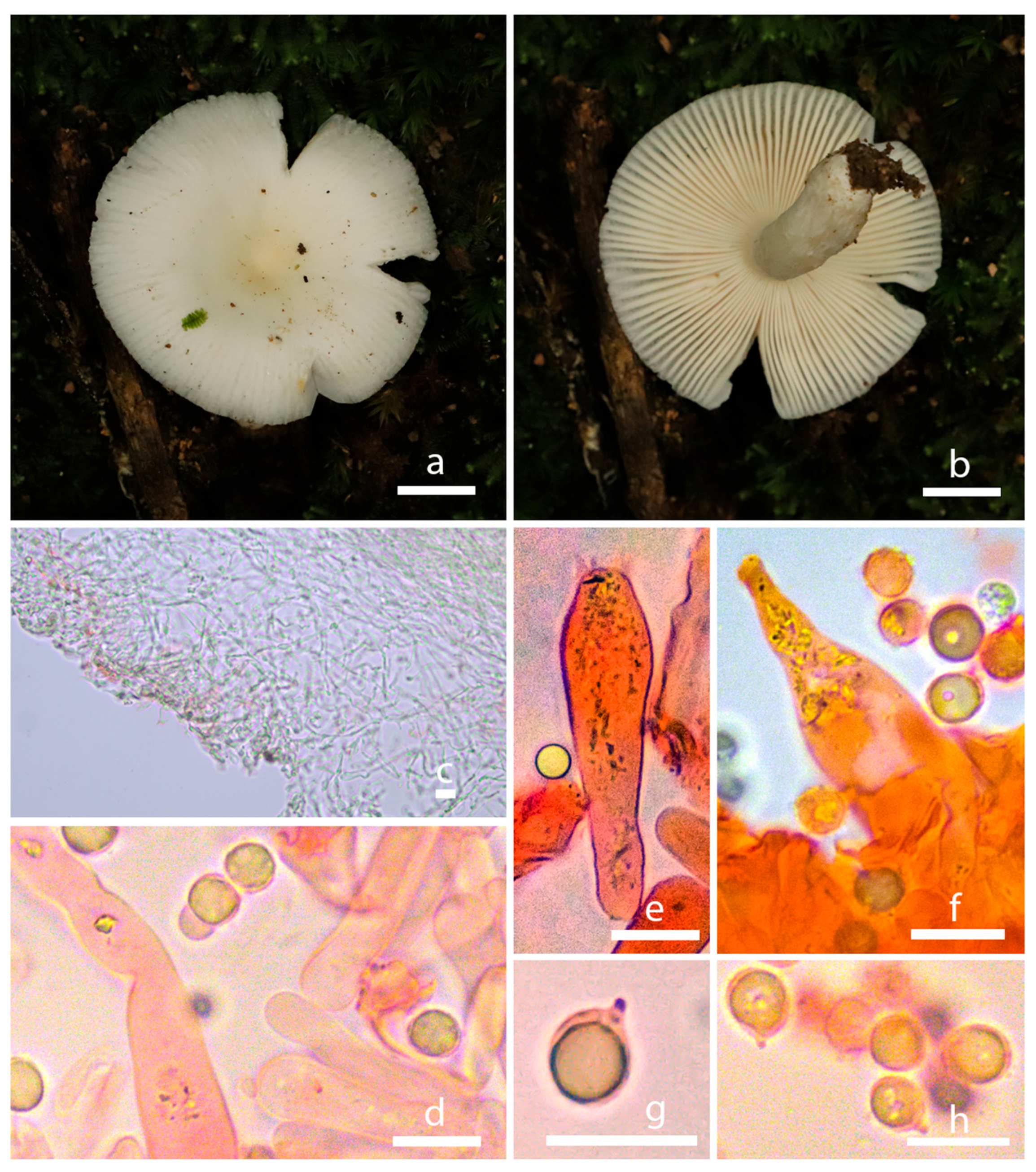
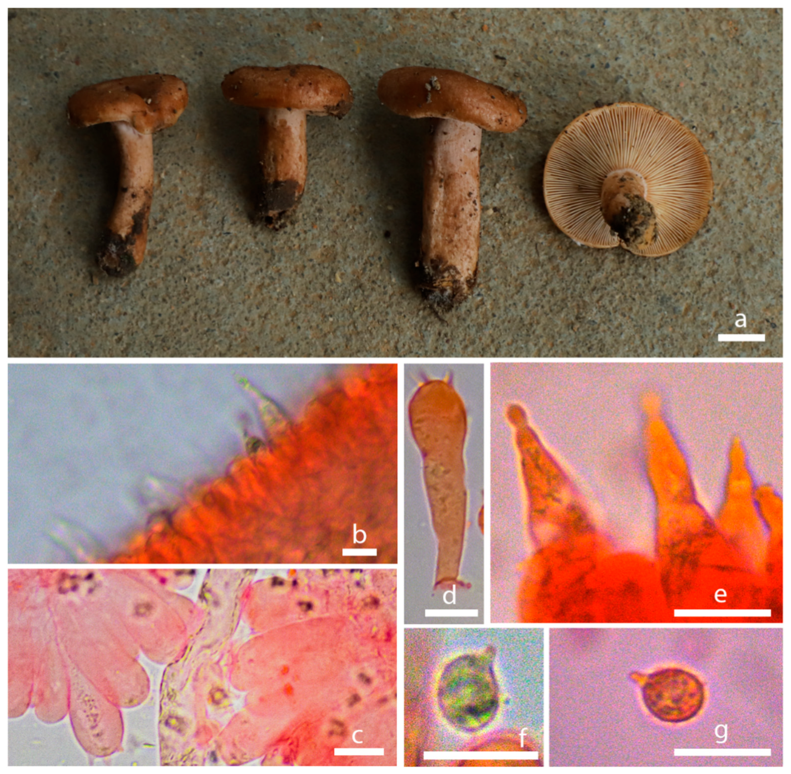
The multi-locus sequence matrix spans a length of 4350 bp. Its dataset comprises 700 bp of ITS, 890 bp of LSU, 1100 bp of TEF1-α, 800 bp of RPB2, and 860 bp of mtSSU. For the multi-locus region, the best substitution model for ITS and RPB2 in the BI analysis is SYM + G4, while for LSU and TEF1-α it is SYM + I + G4, and for mtSSU the best substitution model is GTR + F + G4. A total of 148 sequences, including newly generated ones, were deposited in the GenBank database (Table 1 and Table 2). Based on the foundational rank consistency of the phylogenetic topologies obtained from BI and ML analyses, only the ML trees are depicted in Figure 1 and Figure 2. The resulting phylogenetic trees demonstrate strong support for clades of the four new species in multi-locus phylogenetic analyses. These new species exhibit notable distinctions from known species (Figure 1 and Figure 2). Bootstrap and posterior probability values indicate robust support in multi-locus phylogeny for R. junzifengensis (from subsect. Virescentinae), R. zonatus (from subsect. Virescentinae), and L. jianyangensis (L. subsect. Zonarii), forming a distinct clade.
Lactarius jianyangensis showed the greatest similarity to Lactarius pallido-ochraceus, with an additional 84 sequences from Lactarius collected to construct the tree (Table 1, Figure 1). L. jianyangensis exhibited the highest genetic similarity to L. pallido-ochraceus and clustered with two other species, L. vulgaris and L. pallidizonatus (Figure 1). However, despite clustering with these three species, the substantial phylogenetic distance between L. jianyangensis and the other members of this clade supports its classification as an independent species. Two putative new species within the Russula genus, Russula junzifengensis, formed a strongly supported cluster (BS 100%) and were notably distinct from other known species within the Virescentinae group. Russula junzifengensis clustered together with an unidentified sequence from China (voucher: HMAS250919), which served as the sister clade to R. indoalba, supported by 98% bootstrap support and a posterior probability of 1. R. zonatus clustered alongside R. brunneoaurantiaca and formed a clade sister to R. brunneoaurantiaca with a posterior probability of 1.

3.2. Taxonomy

Russula zonatus S. Liu & Jun Z. Qiu, sp. nov. (Figure 3a,b and Figure 4).
MycoBank: MB 851147.
Etymology: The epithet “zonatus” refers to the morphological feature of ring patterns on the surface.
Holotype: CHINA. Fujian Province, Mingxi County, Xiayang Town, Ziyun Village, in mixed forests, alt. 379 m, 26.34323138° N, 117.45805533° E, 30 August 2021, S. Liu and Jun Z. Qiu (holotype HMAS298100; paratype HMAS298099).
Description: Basidiocarps medium-sized to big. Pileus 7.5–11.9 cm in diam., convex-expanded to infundibuliform with a central depression and slight incurved margin, shallowly infundibuliform when mature, surface glabrous and dry with unclear or golden brown zone lines, no dark brown ring patterns on the surface, brown to grayish brown. Context 3 mm thick, satin white. Lamellae adnate, crowded, marble white, no forking, concolorous with the pileus when fully mature. Stipe 2.5 × 6–3.5 × 10 cm, central, equal, sometimes with fibrils, whitish or sub-concolorous with the pileus.
Basidiospores (6.4) 6.5–7.8 (8) × (5.2) 5.5–6.8 (7) µm (Q = (1.05) 1.06–1.19 (1.23), Q = 1.13 ± 0.05), broadly ellipsoid, with almost isolated warts, plage not amyloid. Basidia 32.5–49 × 10–13 µm, clavate, four-spored. Cystidium common, 30–50 × 5–7.5 µm, fusiform to lanceolate with campulitropal head. Lamella edge sterile, lamellae 8.74 µm thick.
Pileipellis duplex: gelatinous epicutis, 100–230 µm thick, hyphae 2–7 µm wide, hyaline to light yellow in 5% KOH, smooth. Hypodermium well developed, yellowish to lightly yellow intracellular pigment in 5% KOH.
Ecology and distribution: Gregarious in subtropical mixed forests (fagaceous forests or mixed forests with fagaceous trees). Known to inhabit Fujian Province, China.
Russula junzifengensis S. Liu & Jun Z. Qiu, sp. nov. (Figure 3c,d and Figure 5).
MycoBank: MB 851146.
Etymology: Named after the Junzifeng Nature Reserve where the fungus was collected.
Holotype: CHINA. Fujian Province, Mingxi County, Junzifeng Nature Reserve, Xiafang Town, Zhushe Village, in mixed forests, alt. 410 m, 26.56490316° N, 117.03322856° E, 7 August 2021, S. Liu and Jun Z. Qiu (holotype HMAS298101; paratype HMAS298102).
Description: Basidiomata medium-sized, with a diameter of 40–60 mm. Initially hemispherical, later broadly convex to flat with a shallow depression, featuring a sub-transparently striate margin. The lamellae are densely crowded and sometimes slightly decurrent in mature and dry conditions, with a sharp, incurved, and even margin. The surface is glabrous, ranging from dry to slightly glutinous, presenting a satin white appearance, marble white at the center. In the mature stage, the central color turns to a shade of light yellow, pinard yellow, occasionally displaying maize yellow or light orange–yellow to capucine buff. The lamellae are adnate, densely packed, and of yellowish white color, without forking, becoming fragile and matching the pileus color when fully mature. The stipe measures 5.5 × 1.2 cm, central, cylindrical to slightly tapered upwards, rarely becoming subcylindrical to clavate, slightly narrowing towards the base, without an annulus. The stipe is white, appearing yellowish white, smooth in youth, later exhibiting fibrils on the surface. While young, it is full-bodied, eventually becoming hollow. The odor is indistinct.
Basidiospores (5.4) 6–7.5 (7.5) × (5.2) 5.5–6.5 (6.6) µm, [Q = (1.04) 1.05–1.25 (1.25), Q = 1.15 ± 0.06], ellipsoid, composed of small amyloid conical warts, mostly isolated, sometimes fused; indextrinoid, ornamentation small to medium-sized. Basidia 38.5–46.5 × 9–12 µm (smaller), clavate, four-spored, clavate, hyaline to light yellow in 5% KOH. Cystidia, 42 ×13.5 µm (smaller), thin-walled, hyaline in 5% KOH. Pileipellis well developed, separated from the surrounding spherocytes of the context, yellowish to lightly yellow intracellular pigment in 5% KOH.
Ecology and distribution: Gregarious in subtropical mixed forests (solitary or gregarious in Fagaceae forest). Known to inhabit Fujian Province, China.
Lactarius jianyangensis S. Liu & Jun Z. Qiu, sp. nov. (Figure 3e,f and Figure 6).
MycoBank: MB 851149.
Etymology: Named after Jianyang District, where the fungus was collected.
Holotype: CHINA. Fujian Province, Nanping City, Jianyang District, in mixed forests, alt 841 m, 27.34220872° N, 118.16609715° E, 19 August 2021, S. Liu and Jun Z. Qiu (holotype HMAS298103; paratype HMAS298104).
Description: Basidiomata with a small size. Pileus 25–45 mm broad, initially hemispheric, becoming plano-convex and planate when mature, convex with inrolled margin, shallowly infundibuliform when mature, surface greasy when wet, aniline yellow, bittersweet pink, Titian red to agate, sometimes center salmon-orange or Mars yellow with a raw sienna margin, margin glabrous, sub-transparently striate. Context 3–4 mm thick, whitish to brown. The lamella 1–2 mm broad, Mikado orange to cadmium orange when young, xanthine orange, amber brown when mature, concolorous with the pileus when fully mature, sub-crowded to crowded, unequal length and extended. Additionally, the flesh of Lactarius has an aroma. Stipe 35–40 × 7–10 mm, central or tapering downwards, sometimes with longitudinal grooves, surface smooth, greasy, with scattered pits, whitish or sub-concolorous with the pileus, the end of the stipe was slightly enlarged, succulent and hollow, latex white or watery–milky.
Basidiospores (5.99) 6–7.7 (7.8) × (4.8) 4.9–6.3 (6.44) μm, [Q = (1.07) 1.09–1.34 (1.36), Q = 1.20 ± 0.09], broadly ellipsoid, surface has protuberance ridge to reticulate pattern, colorless to hyaline in KOH. Basidia 30.55–39.03 × 8.21–11.32 μm, four-spored, narrowly clavate, colorless to hyaline in KOH, sterigmata 2.84–3.87 μm. Clamp connections abundant in all tissues. The head of pleuromacrocystidia is warping, trama 4.53–4.81 μm.
Ecology and distribution: Gregarious in subtropical fagaceous forests. Known to inhabit Fujian Province, China.

4. Discussion

All Russula and Lactarius species characterized thus far form ectomycorrhizal symbioses with higher plants and trees, and both genera contain cosmopolitan as well as more host-specific members, with both edible and toxic species having been identified [20,38]. Lactarius is characterized by the production of latex, although the genus has now been separated into two (Lactarius and Lactifluus), with an additional separation of several species from Lactarius as well as Russula into Multifurca [39]. Most species of Lactarius form symbioses with broadleaf or coniferous hosts, consistent with their discovery in the Fujian forests of pine.
Russula are distinguished by their bright-colored caps, but do not produce latex and are often characterized by their brittle caps [40]. Due to the difficulty in separating species by their morphological characteristics alone, the modern identification of species within the Russula and Lactarius genera has relied on utilizing the ITS sequence in phylogenetic analysis as the primary molecular method for distinguishing and interpreting these closely related species [41]. However, an overreliance on ITS-based phylogenetic structures can lead to inaccurate subgenus classifications and may overlook the presence of known species, such as the R. queletii complex and the rhodochroa-subsanguinaria complex, often manifested within ITS-based phylogenetics [24]. Additionally, earlier studies focusing on Lactarius species found minimal consistency between Asian Lactarius species and those from other continents. Relying solely on ITS-based phylogenetic analysis and morphological characteristics for Lactarius species’ identification appears insufficient. Hence, the utilization of multi-locus phylogenetic analysis has become the preferred method for revealing the genetic relationships within the Russula and Lactarius genera.
Here, we employed a combined ITS-nrLSU-RBP2-mtSSU-TEF-1α multi-locus phylogenetic analysis method to support the identification of three species that have been named R. junzifengensis, R. zonatus, and L. jianyangensis. These assignments are based on combined morphological characterizations and molecular multilocus phylogenetic analyses. Russula zonatus appears to be a very common red mushroom in the subtropical-tropical Quercus forests of Fujian and is a member of the subgenus Crassotunicata. Key features for its identification include medium basidiocarps, a convex expanded to infundibuliform pileus with a central depression and slight incurved margin, glabrous and dry surface with indistinct or golden brown zone lines, brown to grayish brown color, very crowded lamellae, moderately ornamented basidiospores with isolated warts, and a subtropical habitat. R. zonatus forms a clade with R. brunneoaurantiaca (with a highest ITS identity of ~99%), R. adusta, and R. nigricans. All these species have a mucilaginous pileus and comparatively large spores and basidia. R. zonatus has a high similarity to R.brunneoaurantiaca’s ITS sequence (0.99%), but they have significant morphological differences. R. zonatus belongs to the subgenus Crassotunicata of the Russula genus and has medium to large basidiocarps, with the convex expanded to the infundibuliform pileus with a central depression and slight incurved margin. It has a glabrous and dry surface with indistinct or golden brown zone lines, a brown to grayish brown color, very crowded lamellae, and moderately ornamented basidiospores with isolated warts. R. zonatus has smaller basidia and cystidia compared to R. brunneoaurantiaca, and, at the macroscopic scale, additional differences are quite obvious, with R. brunneoaurantiaca having a surface that is mucilaginous, brownish orange turning yellowish brown to light brown, and a smooth stipe surface [37].
Russula junzifengensis is characterized by a white or slightly stained white–yellow or yellow pileus, which is broadly convex to flat with a shallow depression, slightly crowded lamellae, medium basidiospores with isolated warts, and a subtropical habitat. This species is similar to R. pseudocrustosa, R. indoalba, and R. xanthovirens, and phylogenetic analysis showed that R. junzifengensis formed a highly supported sister group with R. indoalba, but their ITS sequence similarity is less than 90%. Macromorphologically this species seems to be indistinguishable from R. indoalba; both species have whitish gray basidiomata, a clavate stipe, and ellipsoid basidiospores. But the lamellae of R. junzifengensis are not attached to the stipe and appear with fibrils on the stipe.
The phylogenetic results indicate that L. jianyangensis is closest to L. pallido-ochraceus and L. pallidizonatus. In comparison to L. pallido-ochraceus, both species have few pits on their stipe, a surface that is greasy when wet and basidiospores with reticulate ornamentation. In comparison to L. pallido-ochraceus, the basidiomata of the new species described here has a deeper color, smaller basidia, and narrower pleuromacrocystidia. The ITS similarity between the two species was 94%. Southern China’s Lactarius pallidizonatus X.H. Wang seems to be another closely related species [39]; L. pallidizonatus can be distinguished by its light orange–yellow margin, cream–whitish context, pits near the base on stipes, and basidiospores (80/4/3) (7.0) 7.5–9.0 (9.5) × (5.5) 6.0–7.5 µm [Q = (1.10) 1.12–1.29 (1.35), Q = 1.21 ± 0.05], however, L. pallidizonatus has a lighter color and bigger basidiospores than L. jianyangensis.

5. Conclusions

In this study, we employed a multi-locus phylogenetic analysis method, combined with morphological characteristics, to identify three new fungal species in the Quercus forests of Fujian province, namely R. junzifengensis, R. zonatus, and L. jianyangensis. These new species belong to the Russula and Lactarius genera, which form ectomycorrhizal symbioses with higher plants and trees. We found that relying solely on ITS sequences and morphological characteristics for species identification is insufficient, as it may lead to inaccurate subgenus classifications and the omission of known species. Therefore, we suggest using multi-locus phylogenetic analysis methods to reveal the genetic relationships within the Russula and Lactarius genera, as well as their phylogenetic affinities with species from other geographical regions. Our study provides new data on the fungal diversity and distribution in Fujian province, and also contributes new insights to fungal taxonomy and phylogeny.

Author Contributions

Acquisition of funding, Y.C., H.S. and J.Q.; Collection of data, S.L., M.Z., N.O.K., Z.H., J.C., R.C., Y.D., Y.H., Z.W., X.G., C.Y., P.L., W.Z. and H.L. All authors have read and agreed to the published version of the manuscript.

Funding

This research was funded by the National Natural Science Foundation of China (No. 32270029, U1803232, 31670026), the National Key R & D Program of China (No. 2017YFE0122000), a Social Service Team Support Program Project (No. 11899170165), Science and Technology Innovation Special Fund (No. KFB23084) of Fujian Agriculture and Forestry University, a Fujian Provincial Major Science and Technology Project (No. 2022NZ029017), a Key Project from Fujian Provincial Department of Science and Technology (No. 2020N5005), and the Young and Middle-aged Teacher Education Research Project of Fujian Province (No. JAT210075).

Institutional Review Board Statement

Not applicable.

Informed Consent Statement

Not applicable.

Data Availability Statement

All newly generated sequences were deposited in GenBank (https://www.ncbi.nlm.nih.gov/genbank/ (accessed on 10 January 2024)). All new taxa were linked with MycoBank (https://www.mycobank.org/ (accessed on 10 January 2024)).

Acknowledgments

We would like to thank Ling Wang, Longbin Lin, Huili Pu, Tianyu Tan, Taichang Mu, and Jie Zhao for their help with sample collection.

Conflicts of Interest

The authors declare no conflicts of interest.

References

  1. Looney, B.P.; Meidl, P.; Piatek, M.J.; Miettinen, O.; Martin, F.M.; Matheny, P.B.; Labbé, J.L. Russulaceae: A new genomic dataset to study ecosystem function and evolutionary diversification of ectomycorrhizal fungi with their tree associates. New Phytol. 2018, 218, 54–65. [Google Scholar] [CrossRef] [PubMed]
  2. Jiang, X.M.; Li, Y.K.; Liang, J.F.; Wu, J.R. Russula brunneovinacea sp. nov., from Northeastern China. Mycotaxon 2018, 132, 789–797. [Google Scholar] [CrossRef]
  3. Li, G.J.; Zhao, D.; Li, S.F.; Wen, H.A. Russula chiui and R. pseudopectinatoides, two new species from Southwestern China supported by morphological and molecular evidence. Mycol. Prog. 2015, 14, 33. [Google Scholar] [CrossRef]
  4. Taylor, A.F.S.; Alexander, I.J. Ectomycorrhizal synthesis with an isolate of Russula aeruginea. Mycol. Res. 1989, 92, 103–107. [Google Scholar] [CrossRef]
  5. Wang, P.; Zhang, Y.; Mi, F.; Tang, X.; He, X.; Cao, Y.; Liu, C.; Yang, D.; Dong, J.; Zhang, K.; et al. Recent advances in population genetics of ectomycorrhizal mushrooms Russula spp. Mycology 2015, 6, 110–120. [Google Scholar] [CrossRef] [PubMed]
  6. Liu, J.K. Biologically active substances from mushrooms in Yunnan, China. Heterocycles 2002, 57, 157–167. [Google Scholar] [CrossRef]
  7. Singh, R.S.; Walia, A.K.; Kennedy, J.F. Mushroom lectins in biomedical research and development. Int. J. Biol. Macromol. 2020, 151, 1340–1350. [Google Scholar] [CrossRef]
  8. Ali, A.; Guo, D.; Mahar, A.; Wang, P.; Shen, F.; Li, R.H.; Zhang, Z.Q. Mycoremediation of potentially toxic trace elements—A biological tool for soil cleanup: A review. Pedosphere 2017, 27, 205–222. [Google Scholar] [CrossRef]
  9. Chen, F.; Yuan, Y.; Wei, W.; Yu, S.; Zhang, T. Reconstructed Temperature for Yong’an, Fujian, Southeast China: Linkages to the pacific ocean climate variability. Glob. Planet. Chang. 2012, 86–87, 11–19. [Google Scholar] [CrossRef]
  10. Yin, Y.; Gemmer, M.; Luo, Y.; Wang, Y. Tropical cyclones and heavy rainfall in Fujian province, China. Quat. Int. 2010, 226, 122–128. [Google Scholar] [CrossRef]
  11. Liu, Y.; Xu, X.; Dimitrov, D.; Pellissier, L.; Borregaard, M.K.; Shrestha, N.; Su, X.; Luo, A.; Zimmermann, N.E.; Rahbek, C.; et al. An updated floristic map of the world. Nat. Commun. 2023, 14, 2990. [Google Scholar] [CrossRef] [PubMed]
  12. Yue, Y.; Zheng, Z.; Huang, K.; Chevalier, M.; Chase, B.M.; Carré, M.; Ledru, M.P.; Cheddadi, R. A Continuous record of vegetation and climate change over the past 50,000 years in the Fujian province of eastern subtropical China. Palaeogeogr. Palaeoclimatol. Palaeoecol. 2012, 365–366, 115–123. [Google Scholar] [CrossRef]
  13. Wisitrassameewong, K.; Park, M.S.; Lee, H.; Ghosh, A.; Das, K.; Buyck, B.; Looney, B.P.; Caboň, M.; Adamčík, S.; Kim, C.; et al. Taxonomic revision of Russula subsection Amoeninae from South Korea. MycoKeys 2020, 75, 1–29. [Google Scholar] [CrossRef] [PubMed]
  14. Li, G.J.; Dong, Z.; Li, S.F.; Wen, H.A. Recent research progress of Russula (Russulales, Agaricomycetes): A review. Mycosystema 2015, 34, 821–848. [Google Scholar] [CrossRef]
  15. Vera, M.; Adamčík, S.; Adamčíková, K.; Hampe, F.; Caboň, M.; Manz, C.; Ovrebo, C.; Piepenbring, M.; Corrales, A. Morphological and genetic diversification of Russula floriformis, sp. nov., along the Isthmus of Panama. Mycologia 2021, 113, 807–827. [Google Scholar] [CrossRef]
  16. Khatua, S.; Paloi, S.; Acharya, K. An untold story of a novel mushroom from Tribal Cuisine: An Ethno-Medicinal, taxonomic and pharmacological approach. Food Funct. 2021, 12, 4679–4695. [Google Scholar] [CrossRef]
  17. Panda, M.K.; Das, S.K.; Mohapatra, S.; Debata, P.R.; Tayung, K.; Thatoi, H. Mycochemical composition, bioactivities, and phylogenetic placement of three wild edible Russula species from Northern Odisha, India. Plant Biosyst. 2021, 155, 1041–1055. [Google Scholar] [CrossRef]
  18. Looney, B.P.; Manz, C.; Matheny, P.B.; Adamčík, S. Systematic revision of the Roseinae clade of Russula, with a focus on eastern North American taxa. Mycologia 2022, 114, 270–302. [Google Scholar] [CrossRef]
  19. Noffsinger, C.; Cripps, C.L. Systematic analysis of Russula in the North American Rocky Mountain alpine zone. Mycologia 2021, 113, 1278–1315. [Google Scholar] [CrossRef]
  20. Paloi, S.; Kumla, J.; Karunarathna, S.C.; Lumyong, S.; Suwannarach, N. Taxonomic and phylogenetic evidence reveal two new Russula species (Russulaceae, Russulales) from Northern Thailand. Mycol. Prog. 2023, 22, 72. [Google Scholar] [CrossRef]
  21. Miller, S.L.; Buyck, B. Molecular phylogeny of the genus Russula in Europe with a comparison of modern infrageneric classifications. Mycol. Res. 2002, 106, 259–276. [Google Scholar] [CrossRef]
  22. Buyck, B.; Hofstetter, V.; Eberhardt, U.; Verbeken, A.; Kauff, F. Walking the thin line between Russula and Lactarius: The dilemma of Russula subsect. Ochricompactae. Fungal Divers. 2008, 28, 15–40. [Google Scholar]
  23. Buyck, B.; Hofstetter, V.; Verbeken, A.; Walleyn, R. Proposal to conserve Lactarius Nom. Cons. (Basidiomycota) with a conserved type. Taxon 2010, 59, 295–296. [Google Scholar] [CrossRef]
  24. Li, G.J.; Liu, T.Z.; Li, S.M.; Zhao, S.Y.; Niu, C.Y.; Liu, Z.Z.; Xie, X.J.; Zhang, X.; Shi, L.Y.; Guo, Y.B.; et al. Four new species of Russula subsection Sardoninae from China. J. Fungi 2023, 9, 199. [Google Scholar] [CrossRef]
  25. Zhou, H.; Cheng, G.Q.; Wang, Q.T.; Guo, M.J.; Zhuo, L.; Yan, H.F.; Li, G.J.; Hou, C.L. Morphological characteristics and phylogeny reveal six new species in Russula Subgenus Russula (Russulaceae, Russulales) from Yanshan Mountains, North China. J. Fungi 2022, 8, 1283. [Google Scholar] [CrossRef]
  26. Wang, S.H.; Li, G.J.; Phurbu, D.; He, M.Q.; Zhang, M.Z.; Zhu, X.Y.; Li, J.X.; Zhao, R.L.; Cao, B. Four new species of Russula from the Xizang Autonomous Region and other provinces of China. Mycology 2023, 14, 1–28. [Google Scholar] [CrossRef]
  27. Li, G.J.; Li, S.M.; Buyck, B.; Zhao, S.Y.; Xie, X.J.; Shi, L.Y.; Deng, C.Y.; Meng, Q.F.; Sun, Q.B.; Yan, J.Q.; et al. Three new Russula Species in Sect. Ingratae (Russulales, Basidiomycota) from Southern China. MycoKeys 2021, 84, 103–139. [Google Scholar] [CrossRef] [PubMed]
  28. Chen, B.; Song, J.; Liang, J.; Li, Y. Two new species of Russula Subsect. Virescentinae from Southern China. Mycol. Prog. 2021, 20, 993–1005. [Google Scholar] [CrossRef]
  29. Li, F.; Deng, Q.L. Three new species of Russula from South China. Mycol. Prog. 2018, 17, 1305–1321. [Google Scholar] [CrossRef]
  30. White, T.J.; Bruns, T.; Lee, S.; Taylor, J. Amplification and direct sequencing of fungal ribosomal RNA genes for phylogenetics. In PCR Protocols; Elsevier: Amsterdam, The Netherlands, 1990; pp. 315–322. [Google Scholar]
  31. Vilgalys, R.; Hester, M. Rapid genetic identification and mapping of enzymatically amplified ribosomal DNA from several Cryptococcus species. J. Bacteriol. 1990, 172, 4238–4246. [Google Scholar] [CrossRef]
  32. Rehner, S.A.; Buckley, E. A Beauveria phylogeny inferred from nuclear ITS and EF1- sequences: Evidence for cryptic diversification and links to Cordyceps teleomorphs. Mycologia 2005, 97, 84–98. [Google Scholar] [CrossRef] [PubMed]
  33. Liu, Y.J.; Hall, B.D. Body plan evolution of Ascomycetes, as inferred from an RNA polymerase II phylogeny. Proc. Natl. Acad. Sci. USA 2004, 101, 4507–4512. [Google Scholar] [CrossRef] [PubMed]
  34. Chen, B.; Song, J.; Chen, Y.; Zhang, J.; Liang, J. Morphological and phylogenetic evidence for two new species of Russula Subg. Heterophyllidia from Guangdong province of China. MycoKeys 2021, 82, 139–157. [Google Scholar] [CrossRef]
  35. Deng, L.S.; Kang, R.; Zeng, N.K.; Yu, W.J.; Chang, C.; Xu, F.; Deng, W.Q.; Qi, L.L.; Zhou, Y.L.; Fan, Y.G. Two new Inosperma (Inocybaceae) species with unexpected muscarine contents from tropical China. MycoKeys 2021, 85, 87–108. [Google Scholar] [CrossRef]
  36. Buyck, B. One step closer to unravelling the origin of Russula: Subgenus Glutinosae subg. nov. Mycosphere 2020, 11, 285–304. [Google Scholar] [CrossRef]
  37. Roy, N.; Beypih, J.; Tanti, B.; Dutta, A.K. Russula brunneoaurantiaca, a novel taxon of Russula subg. Crassotunicata from west Bengal, India, with morpho-molecular analysis and scanning electron microscopy. Microsc. Res. Tech. 2023, 86, 1–7. [Google Scholar] [CrossRef]
  38. Shi, S.F.; Wang, X.H.; Bau, T. Three new species of Lactarius (Russulaceae, Russulales) from Northeast China. Mycoscience 2018, 59, 206–217. [Google Scholar] [CrossRef]
  39. Wang, X.H. Seven new species of Lactarius subg. Lactarius (Russulaceae) from Southwestern China. Mycosystema 2017, 36, 1463–1482. [Google Scholar] [CrossRef]
  40. Lee, H.; Park, M.S.; Jung, P.E.; Eimes, J.A.; Seok, S.J.; Lim, Y.W. Re-evaluation of the taxonomy and diversity of Russula section Foetentinae (Russulales, Basidiomycota) in Korea. Mycoscience 2017, 58, 351–360. [Google Scholar] [CrossRef]
  41. Adamčík, S.; Looney, B.; Caboň, M.; Jančovičová, S.; Adamčíková, K.; Avis, P.G.; Barajas, M.; Bhatt, R.P.; Corrales, A.; Das, K.; et al. The quest for a globally comprehensible Russula language. Fungal Divers. 2019, 99, 369–449. [Google Scholar] [CrossRef]
Figure 1. Phylogeny inferred from Lactarius multigene sequences (nrLSU, ITS, mtSSU, rpb2, and tef1-α) using Bayesian analysis. Support values in normal type are bootstrap support (BS, significant when ≥70%). Values in bold are Bayesian Posterior Probabilities (PP, significant when ≥0.95). The scale bar indicates the number of nucleotide substitutions per site. New species are highlighted in red. Arrows show the support values at the branching points. Superscript “T” denotes the type strain of the new species.
Jof 10 00070 g001
Figure 2. Phylogeny inferred from Russula multigene sequences (nrLSU, ITS, mtSSU, rpb2, and tef1-α) using Bayesian analysis. Support values in normal type are bootstrap support (BS, significant when ≥70%). Values in bold are Bayesian Posterior Probabilities (PP, significant when ≥0.95). The scale bar indicates the number of nucleotide substitutions per site. New species are highlighted in red. Arrows show the support values at the branching points. Superscript “T” denotes the type strain of the new species.
Jof 10 00070 g002
Figure 3. SEM photos of basidiospores. (a,b) R. zonatus, (c,d) R. junzifengensis, and (e,f) L. jianyangensis. Scale bars: (af) = 10 μm.
Jof 10 00070 g003
Figure 4. Morphological characteristics of Russula zonatus (HMAS298100). (a,b) Basidiomata; (c) pileipellis in 5% KOH; (d) lamellae in 5% KOH. (e,f) Basidium in Congo Red reagent; (g) cystidium in Congo Red reagent; (h) basidiospores in Congo Red reagent; bars: (a,b) = 1 cm; (ch) = 10 µm.
Jof 10 00070 g004
Figure 5. Morphological characteristics of Russula junzifengensis (HMAS298101). (a,b) Basidiomata; (c) pileipellis in 5% KOH; (d) lamellae in Congo Red reagent. (e) Basidium in Congo Red reagent; (f) cystidium in Congo Red reagent; (g,h) basidiospores in Congo Red reagent; bars: (a,b) = 1 cm; (ch) = 10 µm.
Jof 10 00070 g005
Figure 6. Morphological characteristics of Lactarius jianyangensis (HMAS298103). (a) Basidiomata; (b) pileipellis in Congo Red reagent (c) lamellae in Congo Red reagent. (d) Basidium in Congo red reagent; (e) cystidium in Congo red reagent; (f,g) basidiospores in Congo red reagent; bars: (a) = 1 cm; (bg) = 10 µm.
Jof 10 00070 g006
Table 1. Species and specimens of Lactarius used for the molecular phylogenetic analyses.
TaxonVoucherLocationGenBank Accession Number
ITSnrLSURPB2TEF1
L. albocarneus-ChinaKX441117KX441364KX442105-
L. aurantiozonatusHCCN10589KoreaMH984993MH985118MH936950-
L. aurantiozonatusSFC20150901-06KoreaMH984976MH985097MH936929-
L. austroscrobiculatusMHHNU 31161ChinaOL770185---
L. austroscrobiculatusXHW3542ChinaOL770183---
L. blenniusMTB16GermanyMN947353---
L. blennius-SwedenAY606944---
L. brachycystidiatusKUN-HKAS 67933ChinaMF508951---
L. brachycystidiatusKUN-HKAS 96483ChinaMF508950---
L. brachycystidiatusKUN-HKAS 96442ChinaMF508949---
L. brachycystidiatusKUN-HKAS 69233ChinaMF508948---
L. brachycystidiatusKUN-HKAS 76061ChinaMF508947---
L. brachycystidiatusKUN-HKAS69519ChinaMF508952---
L. castaneusKUN-HKAS 47648ChinaMF508962---
L. castaneusKUN-HKAS 61976ChinaMF508961---
L. cinereusLM4663FMexicoFJ348708---
L. cinereusCCB9 (TENN)USAMF755272---
L. cinereus2636CanadaKJ705204---
L. cremicolorKA12-0268KoreaMH985013-QCH40198-
L. cremicolorHCCN19933KoreaMH984972-QCH40153-
L. dilutisalmoneusHMJAU 42465ChinaMF152847---
L. dilutisalmoneusKUN-HKAS 61803ChinaMF152846---
L. fluensIBL83PolandMZ410712---
L. fluensMC01-525DenmarkAJ889961---
L. gloecorpusLH91USAGQ268638---
L. gloeocarpusKUN-HKAS 73602ChinaOL770166---
L. gloeocarpusZP-2287 (MHHNU)ChinaOL770165---
L. gloeocarpusPa1-mOTU056JapanLC315865---
L. haugiaeLM4994MexicoKT583642-AOF41440-
L. haugiaeLM4988MexicoKT583641-AOF41439-
L. haugiaeLM4957MexicoKT583640---
L. hunanensisMHHNU 31989BChinaOL770172---
L. hunanensisMHHNU 31989AChinaOL770171---
L. incrustatusKUN-HKAS 61789ChinaMK675285---
L. incrustatusKUN-HKAS 61772ChinaMK675284---
L. jianyangensisHMAS298103TChinaOR835448OR826782OR915862OR887738
L. jianyangensisHMAS298104ChinaOR835446PP033514OR915863OR887739
L. lignicolaKUN-HKAS 59474ChinaMF508946---
L. lignicolaKUN-HKAS 54122ChinaMF508945---
L. olivinusKUN-HKAS 116928ChinaOL770196---
L. olivinusKUN-HKAS 118988ChinaOL770195---
L. pallidizonatusKUN-HKAS 85389ChinaMF508932---
L. pallidizonatusKUN-HKAS 62019ChinaMF508931---
L. pallidizonatusKUN-HKAS 85388ChinaMF508933---
L. pallidizonatusKUN-HKAS 47641ChinaMF508930---
L. pallido-ochraceusKUN-HKAS 71574ChinaMF508942---
L. pallido-ochraceusKUN-HKAS 54492ChinaMF508941---
L. pallido-ochraceusKUN-HKAS 58675ChinaMF508940---
L. pallido-ochraceusTC_277ChinaMW722813---
L. pallido-ochraceusKUN-HKAS 59446ChinaMF508943---
L. pallido-ochraceusKUN-HKAS 75991ChinaMF508939---
L. parallelusSFC20150701-55KoreaMH984953MH985072MH936904-
L. parallelusKA12-1557KoreaMH984921MH985035MH936867-
L. pohangensisSFC20130807-68KoreaMH985018MH985143MH936975-
L. pohangensisSN1-3870ChinaLC622651---
L. psammicola263825USAMK607513---
L. psammicolaBPL869USAKY848507---
L. purpureocastaneusKUN-HKAS 61975ChinaMF508965---
L. purpureocastaneusKUN-HKAS 56384ChinaMF508964---
L. purpureocastaneusKUN-HKAS 49827ChinaMF508963---
L. scrobiculatusQHU20107ChinaOM970920---
L. scrobiculatusHMAS264940ChinaKX441085KX441332KX442073-
L. scrobiculatusJN01-058ThailandKF432968---
L. scrobiculatusHMAS267747ChinaKX441098 MF893430
L. sinozonariusKUN-HKAS 69414ChinaMF508926---
L. sinozonariusKUN-HKAS 56133ChinaMF508929---
L. sinozonariusKUN-HKAS 48534ChinaMF508928-0-
L. sinozonariusKUN-HKAS 96778ChinaMF508927---
L. sinozonariusKUN-HKAS 54620ChinaMF508925---
L. spadiceusSFC20121005-03KoreaMH985021MH985146MH936978-
L. spadiceusNIBRFG0000104942KoreaMH984956MH985076MH936908-
L. subbrevipesKUN-HKAS 85379ChinaMF508938---
L. subbrevipesKUN-HKAS 61409ChinaMF508937---
L. subbrevipesKUN-HKAS 57562ChinaMF508936---
L. subbrevipesKUN-HKAS 62032ChinaMF508935---
L. subbrevipesKUN-HKAS 94947ChinaMF508934---
L. subzonariusKUN-HKAS 96630ChinaMF508960---
L. subzonariusKUN-HKAS 73522ChinaMF508959---
L. uvidusmh0963SwedenAY606957AF325293--
L. uvidusHMAS269338ChinaKX441140KX441387KX442128-
L. vulgarisMHHNU 32017ChinaOL770178---
L. vulgarisHYJ1878 (EFHAAU 476)ChinaOL770175---
L. vulgarisWNW33 (EFHAAU 1602)ChinaOL770173---
Multifurca zonariaFH12-009ThailandKF432960---
Superscript “T” denotes the type strain of the new species.
Table 2. Species and specimens of Russula used for the molecular phylogenetic analyses.
TaxonVoucherLocationGenBank Accession Number
ITSnrLSURPB2mtSSUTEF1
R. adustaRDL 18-039 BelgiumOM833079---ON015965.1
R. aerugineaAT2003017SwedenDQ421999DQ421999---
R. alboareolataSUT-1ThailandAF345247----
R. alboluteaRITF4460China: Chongqing-MW397121MW411341MW403834-
R. alboluteaRITF4461China: Yunnan-MW397122MW411342MW403835-
R. alboluteaRITF4462China: Yunnan-MW397123MW411343MW403836-
R. alboluteaRITF2653ChinaMT672478MW397120MW411340MW403833-
R. amethystinaSJ84PakistanKT953615----
R. amethystinaSJ92PakistanKT953616----
R. amoenolens12838 (MICH)FranceKF245510----
R. amoenolens77763 (PDD)New ZealandGU222264----
R. ammophilaIC09010703 (BCN)SpainMK112566MK108033---
R. aureoviridisH15060613ChinaKY767808----
R. aureoviridisRITF1868ChinaMW397096--MW403842-
R. aureoviridisH16082612ChinaKY767809MK881920-MK882048MN617846
R. aureoviridisRITF4709ChinaMW646980MW646992-MW647003MW650849
R. brunneoaurantiacaGUBH19957IndiaOP270714----
R. brunneonigraH5813 (DAR)AustraliaEU019945----
R. cerolensHE2720ChinaKC505578----
R. cf. amoenolens12794 (MICH)USAKF245512----
R. cf. vescaBB 06.525Mexico-KU237465KU237751KU237309-
R. crassotunicataUBCOGTR0439sCanadaEU597082----
R. crustosaBPL265USA: Tennessee-KT933826KT933898--
R. cyanoxanthaUE29.09.2002-2France-DQ422033DQ421970--
R. cyanoxanthaFH 12-201GermanyKR364093KR364225---
R. farinipesUE28.09.2002-4SwedenDQ421983----
R. fuliginosaR74CZECH REPUBLICHG798529----
R. griseaBB 07.184Slovakia-KU237509KU237795KU237355-
R. griseaUE2005.08.16-01SwedenDQ422030DQ422030---
R. griseaFH12234GermanyKT934006KT933867---
R. ilicisMF 00.300Italy-KU237595KU237880KU237443-
R. illotaUE26.07.2002-3 (UPS)SwedenDQ422024DQ422024DQ421967--
R. indoalbaAG 15-628IndiaKX234820----
R. junzifengensisHMAS298101TChinaOR826832OR826833OR915864OR941507OR887742
R. junzifengensisHMAS298102ChinaOR880061OR880054OR915865OR941508OR887743
R. langeiBB 07.792France-KU237510KU237796KU237356-
R. livescensF0177ChinaGU371295----
R. maguanensisXHW4765ChinaMH724918MH714537MH939990-MH939983
R. mariaeBB 07.038USA-KU237538KU237824KU237384-
R. medullataBB 07.252Slovakia-KU237546KU237832KU237392-
R. mustelinaFH12226Germany-KT933866KT933937--
R. mustelinaSA 09.88Slovakia-KU237596KU237881KU237444-
R. nigricans-GermanyAF418607----
R. pallescensUBC:F23913CanadaKJ146729----
R. pallidulaRITF2613China-MH027960MH091698MW403845-
R. pallidulaRITF3331China-MH027961MH091699MW403846-
R. parvovirescensSDRM 6280USAMK532789----
R. prasinaHMAS 281232ChinaMH454351----
R. pseudocrustosaHBAU15015ChinaMT337520----
R. pulverulentaRF566 (pers. herb.)USAAY061736----
R. rufobasalisH17052204 ChinaMH168570MK881947.1 -MK882075.1 MT085585.1
R. subpuniceaRITF1435China: Hunan-MW397126MW411346MW403839-
R. subpuniceaRITF2615China: Hunan-MW397127MW411347MW403840-
R. subpuniceaRITF3715ChinaMN833635MW397124MW411344MW403837-
R. subpuniceaRITF2648ChinaMN833638MW397125MW411345MW403838-
R. substriataXHW4766ChinaMH724921MH714540MH939993-MH939986
R. variataBPL241USA-KT933818KT933889--
R. vescaBPL284USAKT933978KT933839---
R. vescaAT2002091SwedenDQ422018DQ422018DQ421959--
R. violeipesBB 07.273Slovakia KU237534KU237820KU237380-
R. virescensHJB9989Belgium DQ422014DQ421955--
R. xanthovirensGDGM 71145ChinaMG786056----
R. zonatusHMAS298099ChinaOR826839OR826846OR915866OR941505OR887740
R. zonatusHMAS298100TChinaOR880062OR880056OR915867OR941506OR887741
Superscript “T” denotes the type strain of the new species.
Disclaimer/Publisher’s Note: The statements, opinions and data contained in all publications are solely those of the individual author(s) and contributor(s) and not of MDPI and/or the editor(s). MDPI and/or the editor(s) disclaim responsibility for any injury to people or property resulting from any ideas, methods, instructions or products referred to in the content.

