
Anatomia 2024, 3(1), 1-7; https://doi.org/10.3390/anatomia3010001 - 03 Jan 2024
Abstract
►
Show Figures
Vertebral and spinal cord anomalies are well known in veterinary medicine. However, nerve root anomalies are seldomly reported. In human patients, nerve root anomalies can cause back pain and radicular pain. In human medicine, nerve root anomalies are more often found in cadaveric
[...] Read more.
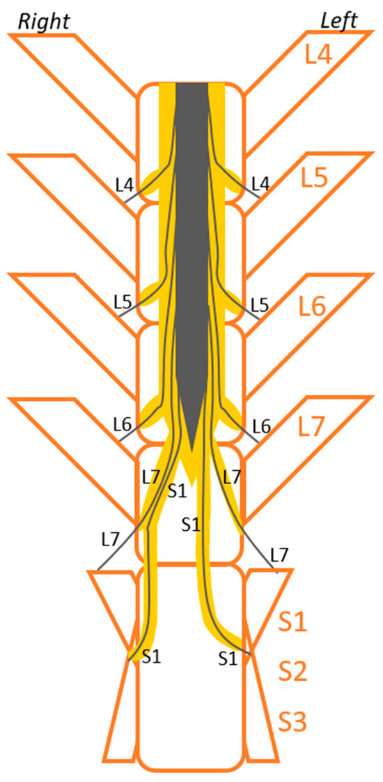
Vertebral and spinal cord anomalies are well known in veterinary medicine. However, nerve root anomalies are seldomly reported. In human patients, nerve root anomalies can cause back pain and radicular pain. In human medicine, nerve root anomalies are more often found in cadaveric studies than in imaging studies, representing the lack of advanced imaging in the past and the unawareness about these pathologies. Additionally, nerve root anomalies can mimic other pathologies in imaging studies. It is important to know about the anatomy of the individual patient not only for correctly localizing the pathology but also for surgical planning and to prevent iatrogenic trauma to the patient. Conjoined nerve roots are a type of nerve root anomaly described in human medicine and are defined as two nerve roots that either share a common dural envelope at some point during their course from the dural sac or that have their origin very close together in the dural sac. In humans, lumbosacral nerve roots are most commonly conjoined, and signs of pain may be associated with this anomaly. We report the magnetic resonance imaging finding of right-sided conjoined L7 and S1 nerve roots in a dog that presented with lumbosacral hyperesthesia. We postulate that it is possible that the conjoined nerve roots played a role in the clinical signs of this dog. This is an anomaly that has not been reported before in veterinary medicine.
Full article
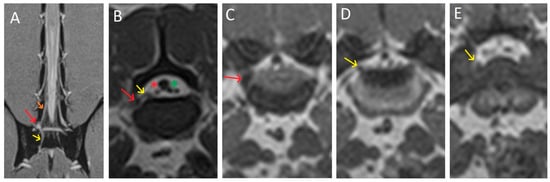
Figure 1


.jpg)


