-
 Reproducibility and Robustness of a Liver Microphysiological System PhysioMimix LC12 under Varying Culture Conditions and Cell Type Combinations
 -
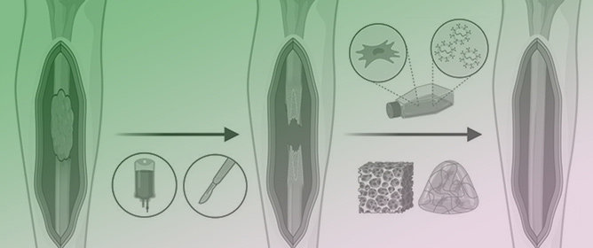 Mesenchymal Stem/Stromal Cells: Immunomodulatory and Bone Regeneration Potential after Tumor Excision in Osteosarcoma Patients
 -
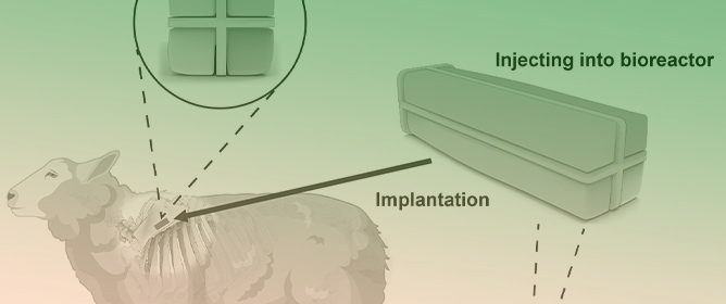 A Comparison of In Vivo Bone Tissue Generation Using Calcium Phosphate Bone Substitutes in a Novel 3D Printed Four-Chamber Periosteal Bioreactor
 -
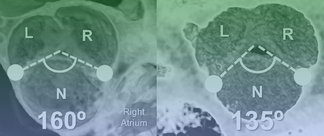 Impact of Variation in Commissural Angle between Fused Leaflets in the Functionally Bicuspid Aortic Valve on Hemodynamics and Tissue Biomechanics
Journal Description
Bioengineering
Bioengineering
is an international, scientific, peer-reviewed, open access journal on the science and technology of bioengineering, published monthly online by MDPI. The Society for Regenerative Medicine (Russian Federation) (RPO) is affiliated with Bioengineering and its members receive discounts on the article processing charges.
- Open Access— free for readers, with article processing charges (APC) paid by authors or their institutions.
- High Visibility: indexed within Scopus, SCIE (Web of Science), PubMed, PMC, CAPlus / SciFinder, Inspec, and other databases.
- Journal Rank: JCR - Q2 (Engineering, Biomedical)
- Rapid Publication: manuscripts are peer-reviewed and a first decision is provided to authors approximately 17.7 days after submission; acceptance to publication is undertaken in 3.6 days (median values for papers published in this journal in the second half of 2023).
- Recognition of Reviewers: reviewers who provide timely, thorough peer-review reports receive vouchers entitling them to a discount on the APC of their next publication in any MDPI journal, in appreciation of the work done.
Impact Factor:
4.6 (2022)
Latest Articles
Reliability of Panoramic Ultrasound in Assessing Rectus Femoris Size, Shape, and Brightness: An Inter-Examiner Study
Bioengineering 2024, 11(1), 82; https://doi.org/10.3390/bioengineering11010082 - 15 Jan 2024
Abstract
Extended field-of-view ultrasound (US) imaging, also known as panoramic US, represents a technical advance that allows for complete visualization of large musculoskeletal structures, which are often limited in conventional 2D US images. Currently, there is no evidence examining whether the experience of examiners
[...] Read more.
Extended field-of-view ultrasound (US) imaging, also known as panoramic US, represents a technical advance that allows for complete visualization of large musculoskeletal structures, which are often limited in conventional 2D US images. Currently, there is no evidence examining whether the experience of examiners influences muscle shape deformations that may arise during the glide of the transducer in panoramic US acquisition. As no studies using panoramic US have analyzed whether two examiners with differing levels of experience might obtain varying scores in size, shape, or brightness during the US assessment of the rectus femoris muscle, our aim was to analyze the inter-examiner reliability of panoramic US imaging acquisition in determining muscle size, shape, and brightness between two examiners. Additionally, we sought to investigate whether the examiners’ experience plays a significant role in muscle deformations during imaging acquisition by assessing score differences. Shape (circularity, aspect ratio, and roundness), size (cross-sectional area and perimeter), and brightness (mean echo intensity) were analyzed in 39 volunteers. Intraclass correlation coefficients (ICCs), standard error of measurements (SEM), minimal detectable changes (MDC), and coefficient of absolute errors (CAE%) were calculated. All parameters evaluated showed no significant differences between the two examiners (p > 0.05). Panoramic US proved to be reliable, regardless of examiner experience, as no deformations were observed. Further research is needed to corroborate the validity of panoramic US by comparing this method with gold standard techniques.
Full article
(This article belongs to the Special Issue New Insights into Photoacoustic Imaging in Biomedicine: Updates and Directions)
►
Show Figures
Open AccessArticle
Implementation of Fluorescent-Protein-Based Quantification Analysis in L-Form Bacteria
Bioengineering 2024, 11(1), 81; https://doi.org/10.3390/bioengineering11010081 - 15 Jan 2024
Abstract
Cell-wall-less (L-form) bacteria exhibit morphological complexity and heterogeneity, complicating quantitative analysis of them under internal and external stimuli. Stable and efficient labeling is needed for the fluorescence-based quantitative cell analysis of L-forms during growth and proliferation. Here, we evaluated the expression of multiple
[...] Read more.
Cell-wall-less (L-form) bacteria exhibit morphological complexity and heterogeneity, complicating quantitative analysis of them under internal and external stimuli. Stable and efficient labeling is needed for the fluorescence-based quantitative cell analysis of L-forms during growth and proliferation. Here, we evaluated the expression of multiple fluorescent proteins (FPs) under different promoters in the Bacillus subtilis L-form strain LR2 using confocal microscopy and imaging flow cytometry. Among others, Pylb-derived NBP3510 showed a superior performance for inducing several FPs including EGFP and mKO2 in both the wild-type and L-form strains. Moreover, NBP3510 was also active in Escherichia coli and its L-form strain NC-7. Employing these established FP-labeled strains, we demonstrated distinct morphologies in the L-form bacteria in a quantitative manner. Given cell-wall-deficient bacteria are considered protocell and synthetic cell models, the generated cell lines in our work could be valuable for L-form-based research.
Full article
(This article belongs to the Section Cellular and Molecular Bioengineering)
►▼
Show Figures
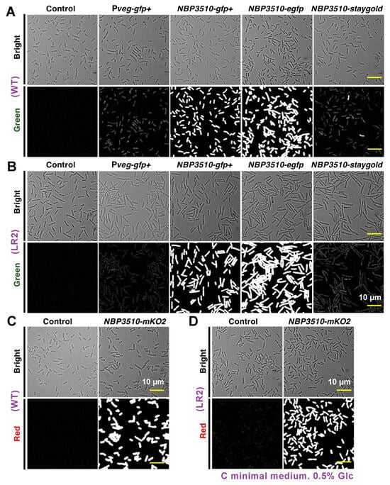
Figure 1
Open AccessArticle
Features of Vat-Photopolymerized Masters for Microfluidic Device Manufacturing
by
, , , , , , and
Bioengineering 2024, 11(1), 80; https://doi.org/10.3390/bioengineering11010080 - 15 Jan 2024
Abstract
The growing interest in advancing microfluidic devices for manipulating fluids within micrometer-scale channels has prompted a shift in manufacturing practices, moving from single-component production to medium-size batches. This transition arises due to the impracticality of lab-scale manufacturing methods in accommodating the increased demand.
[...] Read more.
The growing interest in advancing microfluidic devices for manipulating fluids within micrometer-scale channels has prompted a shift in manufacturing practices, moving from single-component production to medium-size batches. This transition arises due to the impracticality of lab-scale manufacturing methods in accommodating the increased demand. This experimental study focuses on the design of master benchmarks 1–5, taking into consideration critical parameters such as rib width, height, and the relative width-to-height ratio. Notably, benchmarks 4 and 5 featured ribs that were strategically connected to the inlet, outlet, and reaction chamber of the master, enhancing their utility for subsequent replica production. Vat photopolymerization was employed for the fabrication of benchmarks 1–5, while replicas of benchmarks 4 and 5 were generated through polydimethylsiloxane casting. Dimensional investigations of the ribs and channels in both the master benchmarks and replicas were conducted using an optical technique validated through readability analysis based on the Michelson global contrast index. The primary goal was to evaluate the potential applicability of vat photopolymerization technology for efficiently producing microfluidic devices through a streamlined production process. Results indicate that the combination of vat photopolymerization followed by replication is well suited for achieving a minimum rib size of 25 µm in width and an aspect ratio of 1:12 for the master benchmark.
Full article
(This article belongs to the Special Issue Microfluidics and Sensor Technology in Biomedical Engineering)
►▼
Show Figures
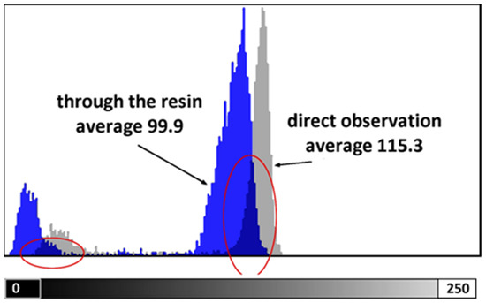
Figure 1
Open AccessArticle
COVID-19 Detection and Diagnosis Model on CT Scans Based on AI Techniques
Bioengineering 2024, 11(1), 79; https://doi.org/10.3390/bioengineering11010079 - 14 Jan 2024
Abstract
The end of 2019 could be mounted in a rudimentary framing of a new medical problem, which globally introduces into the discussion a fulminant outbreak of coronavirus, consequently spreading COVID-19 that conducted long-lived and persistent repercussions. Hence, the theme proposed to be solved
[...] Read more.
The end of 2019 could be mounted in a rudimentary framing of a new medical problem, which globally introduces into the discussion a fulminant outbreak of coronavirus, consequently spreading COVID-19 that conducted long-lived and persistent repercussions. Hence, the theme proposed to be solved arises from the field of medical imaging, where a pulmonary CT-based standardized reporting system could be addressed as a solution. The core of it focuses on certain impediments such as the overworking of doctors, aiming essentially to solve a classification problem using deep learning techniques, namely, if a patient suffers from COVID-19, viral pneumonia, or is healthy from a pulmonary point of view. The methodology’s approach was a meticulous one, denoting an empirical character in which the initial stage, given using data processing, performs an extraction of the lung cavity from the CT scans, which is a less explored approach, followed by data augmentation. The next step is comprehended by developing a CNN in two scenarios, one in which there is a binary classification (COVID and non-COVID patients), and the other one is represented by a three-class classification. Moreover, viral pneumonia is addressed. To obtain an efficient version, architectural changes were gradually made, involving four databases during this process. Furthermore, given the availability of pre-trained models, the transfer learning technique was employed by incorporating the linear classifier from our own convolutional network into an existing model, with the result being much more promising. The experimentation encompassed several models including MobileNetV1, ResNet50, DenseNet201, VGG16, and VGG19. Through a more in-depth analysis, using the CAM technique, MobilneNetV1 differentiated itself via the detection accuracy of possible pulmonary anomalies. Interestingly, this model stood out as not being among the most used in the literature. As a result, the following values of evaluation metrics were reached: loss (0.0751), accuracy (0.9744), precision (0.9758), recall (0.9742), AUC (0.9902), and F1 score (0.9750), from 1161 samples allocated for each of the three individual classes.
Full article
(This article belongs to the Special Issue Artificial Intelligence in Biomedical Diagnosis and Prognosis)
►▼
Show Figures

Graphical abstract
Open AccessArticle
Photobiomodulation Therapy Improves Repair of Bone Defects Filled by Inorganic Bone Matrix and Fibrin Heterologous Biopolymer
by
, , , , , , , , and
Bioengineering 2024, 11(1), 78; https://doi.org/10.3390/bioengineering11010078 - 13 Jan 2024
Abstract
Biomaterials are used extensively in graft procedures to correct bone defects, interacting with the body without causing adverse reactions. The aim of this pre-clinical study was to analyze the effects of photobiomodulation therapy (PBM) with the use of a low-level laser in the
[...] Read more.
Biomaterials are used extensively in graft procedures to correct bone defects, interacting with the body without causing adverse reactions. The aim of this pre-clinical study was to analyze the effects of photobiomodulation therapy (PBM) with the use of a low-level laser in the repair process of bone defects filled with inorganic matrix (IM) associated with heterologous fibrin biopolymer (FB). A circular osteotomy of 4 mm in the left tibia was performed in 30 Wistar male adult rats who were randomly divided into three groups: G1 = IM + PBM, G2 = IM + FB and G3 = IM + FB + PBM. PBM was applied at the time of the experimental surgery and three times a week, on alternate days, until euthanasia, with 830 nm wavelength, in two points of the operated site. Five animals from each group were euthanized 14 and 42 days after surgery. In the histomorphometric analysis, the percentage of neoformed bone tissue in G3 (28.4% ± 2.3%) was higher in relation to G1 (24.1% ± 2.91%) and G2 (22.2% ± 3.11%) at 14 days and at 42 days, the percentage in G3 (35.1% ± 2.55%) was also higher in relation to G1 (30.1% ± 2.9%) and G2 (31.8% ± 3.12%). In the analysis of the birefringence of collagen fibers, G3 showed a predominance of birefringence between greenish-yellow in the neoformed bone tissue after 42 days, differing from the other groups with a greater presence of red-orange fibers. Immunohistochemically, in all experimental groups, it was possible to observe immunostaining for osteocalcin (OCN) near the bone surface of the margins of the surgical defect and tartrate-resistant acid phosphatase (TRAP) bordering the newly formed bone tissue. Therefore, laser photobiomodulation therapy contributed to improving the bone repair process in tibial defects filled with bovine biomaterial associated with fibrin biopolymer derived from snake venom.
Full article
(This article belongs to the Special Issue Biomaterials for Cartilage and Bone Tissue Engineering)
►▼
Show Figures
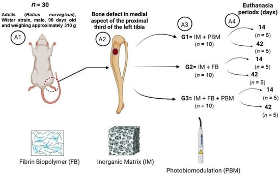
Figure 1
Open AccessArticle
Optimizing RNNs for EMG Signal Classification: A Novel Strategy Using Grey Wolf Optimization
by
, , , and
Bioengineering 2024, 11(1), 77; https://doi.org/10.3390/bioengineering11010077 - 13 Jan 2024
Abstract
Accurate classification of electromyographic (EMG) signals is vital in biomedical applications. This study evaluates different architectures of recurrent neural networks for the classification of EMG signals associated with five movements of the right upper extremity. A Butterworth filter was implemented for signal preprocessing,
[...] Read more.
Accurate classification of electromyographic (EMG) signals is vital in biomedical applications. This study evaluates different architectures of recurrent neural networks for the classification of EMG signals associated with five movements of the right upper extremity. A Butterworth filter was implemented for signal preprocessing, followed by segmentation into 250 ms windows, with an overlap of 190 ms. The resulting dataset was divided into training, validation, and testing subsets. The Grey Wolf Optimization algorithm was applied to the gated recurrent unit (GRU), long short-term memory (LSTM) architectures, and bidirectional recurrent neural networks. In parallel, a performance comparison with support vector machines (SVMs) was performed. The results obtained in the first experimental phase revealed that all the RNN networks evaluated reached a 100% accuracy, standing above the 93% achieved by the SVM. Regarding classification speed, LSTM ranked as the fastest architecture, recording a time of 0.12 ms, followed by GRU with 0.134 ms. Bidirectional recurrent neural networks showed a response time of 0.2 ms, while SVM had the longest time at 2.7 ms. In the second experimental phase, a slight decrease in the accuracy of the RNN models was observed, standing at 98.46% for LSTM, 96.38% for GRU, and 97.63% for the bidirectional network. The findings of this study highlight the effectiveness and speed of recurrent neural networks in the EMG signal classification task.
Full article
(This article belongs to the Special Issue Bioengineering of the Motor System)
►▼
Show Figures
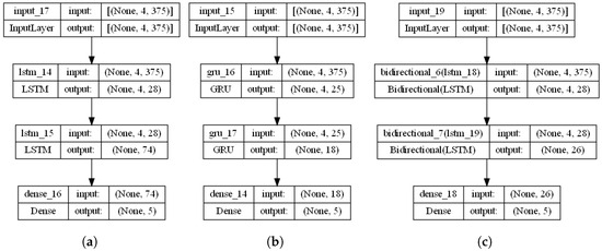
Figure 1
Open AccessArticle
Flow Diverter Performance Comparison of Different Wire Materials for Effective Intracranial Aneurysm Treatment
Bioengineering 2024, 11(1), 76; https://doi.org/10.3390/bioengineering11010076 - 12 Jan 2024
Abstract
A flow diverter (FD) is an effective method for treating wide-necked intracranial aneurysms by inducing hemodynamic changes in aneurysms. However, the procedural technique remains challenging, and it is often not performed properly in many cases of deployment or placements. In this study, three
[...] Read more.
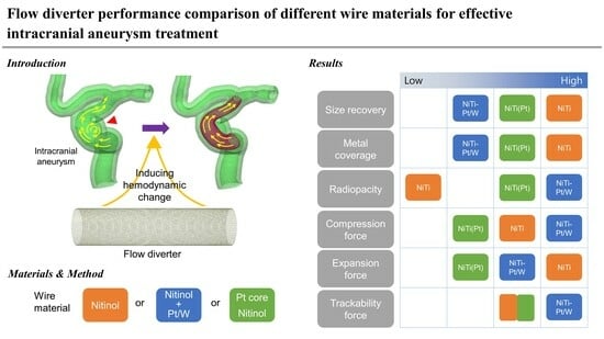
A flow diverter (FD) is an effective method for treating wide-necked intracranial aneurysms by inducing hemodynamic changes in aneurysms. However, the procedural technique remains challenging, and it is often not performed properly in many cases of deployment or placements. In this study, three types of FDs that changed the material of the wire were prepared within the same structure. Differences in physical properties, such as before and after delivery loading stent size, radial force, and radiopacity, were evaluated. The performances in terms of deployment and trackability force were also evaluated in a simulated model using these FDs. Furthermore, changes of deployment patterns when these FDs were applied to a 3D-printed aneurysm model were determined. The NiTi FD using only nitinol (NiTi) wire showed 100% size recovery and 42% to 45% metal coverage after loading. The low trackability force (10.9 to 22.9 gf) allows smooth movement within the delivery system. However, NiTi FD cannot be used in actual surgeries due to difficulties in X-ray identification. NiTi-Pt/W FD, a combination of NiTi wire and platinum/tungsten (Pt/W) wire, had the highest radiopacity and compression force (6.03 ± 0.29 gf) among the three FDs. However, it suffered from high trackability force (22.4 to 39.9 gf) and the end part braiding mesh tended to loosen easily, so the procedure became more challenging. The NiTi(Pt) FD using a platinum core nitinol (NiTi(Pt)) wire had similar trackability force (11.3 to 22.1 gf) to NiTi FD and uniform deployment, enhancing procedural convenience. However, concerns about low expansion force (1.79 ± 0.30 gf) and the potential for migration remained. This comparative analysis contributes to a comprehensive understanding of how different wire materials influence the performance of FDs. While this study is still in its early stages and requires further research, its development has the potential to guide clinicians and researchers in optimizing the selection and development of FDs for the effective treatment of intracranial aneurysms.
Full article
(This article belongs to the Special Issue Using Existing Medical Technologies to Solve and Improve Medical Challenges)
►▼
Show Figures
Graphical abstract
Open AccessArticle
Feasibility of a Shape-Memory-Alloy-Actuator System for Modular Acetabular Cups
by
, , , , and
Bioengineering 2024, 11(1), 75; https://doi.org/10.3390/bioengineering11010075 - 12 Jan 2024
Abstract
Hip implants have a modular structure which enables patient-specific adaptation but also revision of worn or damaged friction partners without compromising the implant-bone connection. To reduce complications during the extraction of ceramic inlays, this work presents a new approach of a shape-memory-alloy-actuator which
[...] Read more.
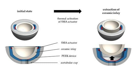
Hip implants have a modular structure which enables patient-specific adaptation but also revision of worn or damaged friction partners without compromising the implant-bone connection. To reduce complications during the extraction of ceramic inlays, this work presents a new approach of a shape-memory-alloy-actuator which enables the loosening of ceramic inlays from acetabular hip cups without ceramic chipping or damaging the metal cup. This technical in vitro study exam-ines two principles of heating currents and hot water for thermal activation of the shape-memory-alloy-actuator to generate a force between the metal cup and the ceramic inlay. Mechanical tests concerning push-in and push-out forces, deformation of the acetabular cup according to international test standards, and force generated by the actuator were generated to prove the feasibility of this new approach to ceramic inlay revision. The required disassembly force for a modular acetabular device achieved an average value of 602 N after static and 713 N after cyclic loading. The actuator can provide a push-out force up to 1951 N. In addition, it is shown that the necessary modifications to the implant modules for the implementation of the shape-memory-actuator-system do not result in any change in the mechanical properties compared to conventional systems.
Full article
(This article belongs to the Special Issue Innovative Medical Technology and Surgical Techniques: Focus on Joint Arthroplasty)
►▼
Show Figures
Graphical abstract
Open AccessArticle
Osteoblast Attachment on Bioactive Glass Air Particle Abrasion-Induced Calcium Phosphate Coating
by
, , , , , and
Bioengineering 2024, 11(1), 74; https://doi.org/10.3390/bioengineering11010074 - 12 Jan 2024
Abstract
Air particle abrasion (APA) using bioactive glass (BG) effectively decontaminates titanium (Ti) surface biofilms and the retained glass particles on the abraded surfaces impart potent antibacterial properties against various clinically significant pathogens. The objective of this study was to investigate the effect of
[...] Read more.
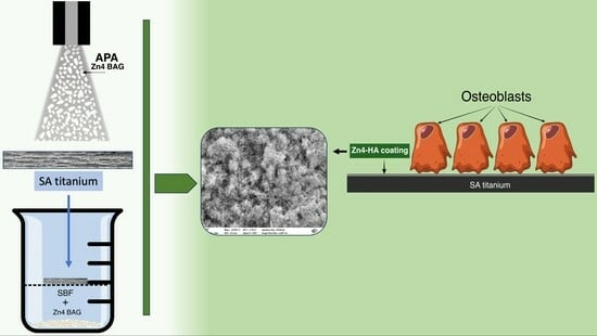
Air particle abrasion (APA) using bioactive glass (BG) effectively decontaminates titanium (Ti) surface biofilms and the retained glass particles on the abraded surfaces impart potent antibacterial properties against various clinically significant pathogens. The objective of this study was to investigate the effect of BG APA and simulated body fluid (SBF) immersion of sandblasted and acid-etched (SA) Ti surfaces on osteoblast cell viability. Another goal was to study the antibacterial effect against Streptococcus mutans. Square-shaped 10 mm diameter Ti substrates (n = 136) were SA by grit blasting with aluminum oxide particles, then acid-etching in an HCl-H2SO4 mixture. The SA substrates (n = 68) were used as non-coated controls (NC-SA). The test group (n = 68) was further subjected to APA using experimental zinc-containing BG (Zn4) and then mineralized in SBF for 14 d (Zn4-CaP). Surface roughness, contact angle, and surface free energy (SFE) were calculated on test and control surfaces. In addition, the topography and chemistry of substrate surfaces were also characterized. Osteoblastic cell viability and focal adhesion were also evaluated and compared to glass slides as an additional control. The antibacterial effect of Zn4-CaP was also assessed against S. mutans. After immersion in SBF, a mineralized zinc-containing Ca-P coating was formed on the SA substrates. The Zn4-CaP coating resulted in a significantly lower Ra surface roughness value (2.565 μm; p < 0.001), higher wettability (13.35°; p < 0.001), and higher total SFE (71.13; p < 0.001) compared to 3.695 μm, 77.19° and 40.43 for the NC-SA, respectively. APA using Zn4 can produce a zinc-containing calcium phosphate coating that demonstrates osteoblast cell viability and focal adhesion comparable to that on NC-SA or glass slides. Nevertheless, the coating had no antibacterial effect against S. mutans.
Full article
(This article belongs to the Special Issue Titanium Implant and Its Cleaning/Decontamination Techniques)
►▼
Show Figures
Graphical abstract
Open AccessArticle
Data-Driven Insights into Labor Progression with Gaussian Processes
Bioengineering 2024, 11(1), 73; https://doi.org/10.3390/bioengineering11010073 - 11 Jan 2024
Abstract
Clinicians routinely perform pelvic examinations to assess the progress of labor. Clinical guidelines to interpret these examinations, using time-based models of cervical dilation, are not always followed and have not contributed to reducing cesarean-section rates. We present a novel Gaussian process model of
[...] Read more.
Clinicians routinely perform pelvic examinations to assess the progress of labor. Clinical guidelines to interpret these examinations, using time-based models of cervical dilation, are not always followed and have not contributed to reducing cesarean-section rates. We present a novel Gaussian process model of labor progress, suitable for real-time use, that predicts cervical dilation and fetal station based on clinically relevant predictors available from the pelvic exam and cardiotocography. We show that the model is more accurate than a statistical approach using a mixed-effects model. In addition, it provides confidence estimates on the prediction, calibrated to the specific delivery. Finally, we show that predicting both dilation and station with a single Gaussian process model is more accurate than two separate models with single predictions.
Full article
(This article belongs to the Special Issue Fetal-Maternal Monitoring during Pregnancy and Labor: Trends and Opportunities)
►▼
Show Figures

Figure 1
Open AccessReview
Multi-Dimensional Modeling of Cerebral Hemodynamics: A Systematic Review
Bioengineering 2024, 11(1), 72; https://doi.org/10.3390/bioengineering11010072 - 11 Jan 2024
Abstract
The Circle of Willis (CoW) describes the arterial system in the human brain enabling the neurovascular blood supply. Neurovascular diseases like intracranial aneurysms (IAs) can occur within the CoW and carry the risk of rupture, which can lead to subarachnoid hemorrhage. The assessment
[...] Read more.
The Circle of Willis (CoW) describes the arterial system in the human brain enabling the neurovascular blood supply. Neurovascular diseases like intracranial aneurysms (IAs) can occur within the CoW and carry the risk of rupture, which can lead to subarachnoid hemorrhage. The assessment of hemodynamic information in these pathologies is crucial for their understanding regarding detection, diagnosis and treatment. Multi-dimensional in silico approaches exist to evaluate these hemodynamics based on patient-specific input data. The approaches comprise low-scale (zero-dimensional, one-dimensional) and high-scale (three-dimensional) models as well as multi-scale coupled models. The input data can be derived from medical imaging, numerical models, literature-based assumptions or from measurements within healthy subjects. Thus, the most realistic description of neurovascular hemodynamics is still controversial. Within this systematic review, first, the models of the three scales (0D, 1D, 3D) and second, the multi-scale models, which are coupled versions of the three scales, were discussed. Current best practices in describing neurovascular hemodynamics most realistically and their clinical applicablility were elucidated. The performance of 3D simulation entails high computational expenses, which could be reduced by analyzing solely the region of interest in detail. Medical imaging to establish patient-specific boundary conditions is usually rare, and thus, lower dimensional models provide a realistic mimicking of the surrounding hemodynamics. Multi-scale coupling, however, is computationally expensive as well, especially when taking all dimensions into account. In conclusion, the 0D–1D–3D multi-scale approach provides the most realistic outcome; nevertheless, it is least applicable. A 1D–3D multi-scale model can be considered regarding a beneficial trade-off between realistic results and applicable performance.
Full article
(This article belongs to the Section Regenerative Engineering)
►▼
Show Figures
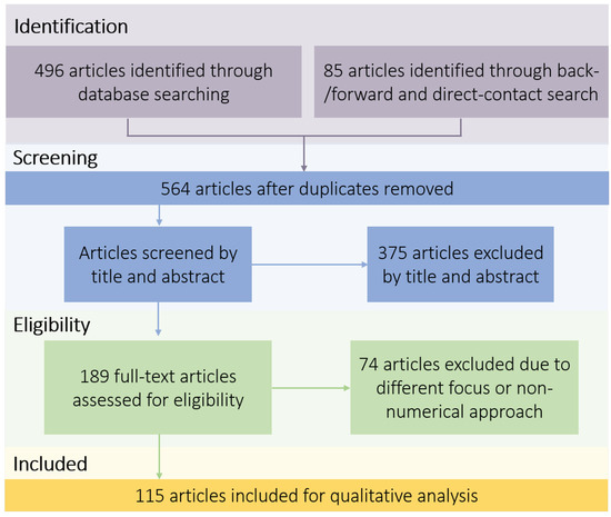
Figure 1
Open AccessArticle
U-Net Convolutional Neural Network for Real-Time Prediction of the Number of Cultured Corneal Endothelial Cells for Cellular Therapy
by
, , , , , , , and
Bioengineering 2024, 11(1), 71; https://doi.org/10.3390/bioengineering11010071 - 11 Jan 2024
Abstract
Corneal endothelial decompensation is treated by the corneal transplantation of donor corneas, but donor shortages and other problems associated with corneal transplantation have prompted investigations into tissue engineering therapies. For clinical use, cells used in tissue engineering must undergo strict quality control to
[...] Read more.
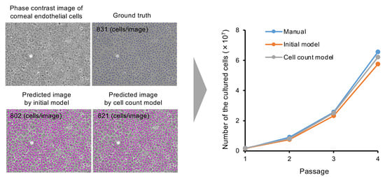
Corneal endothelial decompensation is treated by the corneal transplantation of donor corneas, but donor shortages and other problems associated with corneal transplantation have prompted investigations into tissue engineering therapies. For clinical use, cells used in tissue engineering must undergo strict quality control to ensure their safety and efficacy. In addition, efficient cell manufacturing processes are needed to make cell therapy a sustainable standard procedure with an acceptable economic burden. In this study, we obtained 3098 phase contrast images of cultured human corneal endothelial cells (HCECs). We labeled the images using semi-supervised learning and then trained a model that predicted the cell centers with a precision of 95.1%, a recall of 92.3%, and an F-value of 93.4%. The cell density calculated by the model showed a very strong correlation with the ground truth (Pearson’s correlation coefficient = 0.97, p value = 8.10 × 10−52). The total cell numbers calculated by our model based on phase contrast images were close to the numbers calculated using a hemocytometer through passages 1 to 4. Our findings confirm the feasibility of using artificial intelligence-assisted quality control assessments in the field of regenerative medicine.
Full article
(This article belongs to the Special Issue New Strategies to Treat Corneal Endothelial Diseases: Cell Therapy, Bioengineering of Corneal Grafts and Pharmacological Treatment)
►▼
Show Figures
Graphical abstract
Open AccessArticle
DSP-KD: Dual-Stage Progressive Knowledge Distillation for Skin Disease Classification
Bioengineering 2024, 11(1), 70; https://doi.org/10.3390/bioengineering11010070 - 10 Jan 2024
Abstract
The increasing global demand for skin disease diagnostics emphasizes the urgent need for advancements in AI-assisted diagnostic technologies for dermatoscopic images. In current practical medical systems, the primary challenge is balancing lightweight models with accurate image analysis to address constraints like limited storage
[...] Read more.
The increasing global demand for skin disease diagnostics emphasizes the urgent need for advancements in AI-assisted diagnostic technologies for dermatoscopic images. In current practical medical systems, the primary challenge is balancing lightweight models with accurate image analysis to address constraints like limited storage and computational costs. While knowledge distillation methods hold immense potential in healthcare applications, related research on multi-class skin disease tasks is scarce. To bridge this gap, our study introduces an enhanced multi-source knowledge fusion distillation framework, termed DSP-KD, which improves knowledge transfer in a dual-stage progressive distillation approach to maximize mutual information between teacher and student representations. The experimental results highlight the superior performance of our distilled ShuffleNetV2 on both the ISIC2019 dataset and our private skin disorders dataset. Compared to other state-of-the-art distillation methods using diverse knowledge sources, the DSP-KD demonstrates remarkable effectiveness with a smaller computational burden.
Full article
(This article belongs to the Special Issue Artificial Intelligence for Better Healthcare and Precision Medicine)
►▼
Show Figures
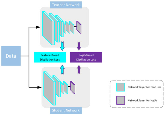
Figure 1
Open AccessArticle
Intraoperative Laparoscopic Hyperspectral Imaging during Esophagectomy—A Pilot Study Evaluating Esophagogastric Perfusion at the Anastomotic Sites
by
, , , , , , and
Bioengineering 2024, 11(1), 69; https://doi.org/10.3390/bioengineering11010069 - 09 Jan 2024
Abstract
Hyperspectral imaging (HSI) is a non-invasive and contactless technique that enables the real-time acquisition of comprehensive information on tissue within the surgical field. In this pilot study, we investigated whether a new HSI system for minimally-invasive surgery, TIVITA® Mini (HSI-MIS), provides reliable
[...] Read more.
Hyperspectral imaging (HSI) is a non-invasive and contactless technique that enables the real-time acquisition of comprehensive information on tissue within the surgical field. In this pilot study, we investigated whether a new HSI system for minimally-invasive surgery, TIVITA® Mini (HSI-MIS), provides reliable insights into tissue perfusion of the proximal and distal esophagogastric anastomotic sites during 21 laparoscopic/thoracoscopic or robotic Ivor Lewis esophagectomies of patients with cancer to minimize the risk of dreaded anastomotic insufficiency. In this pioneering investigation, physiological tissue parameters were derived from HSI measurements of the proximal site of the anastomosis (esophageal stump) and the distal site of the anastomosis (tip of the gastric conduit) during the thoracic phase of the procedure. Tissue oxygenation (StO2), Near Infrared Perfusion Index (NIR-PI), and Tissue Water Index (TWI) showed similar median values at both anastomotic sites. Significant differences were observed only for NIR-PI (median: 76.5 vs. 63.9; p = 0.012) at the distal site (gastric conduit) compared to our previous study using an HSI system for open surgery. For all 21 patients, reliable and informative measurements were attainable, confirming the feasibility of HSI-MIS to assess anastomotic viability. Further studies on the added benefit of this new technique aiming to reduce anastomotic insufficiency are warranted.
Full article
(This article belongs to the Special Issue Application of Hyperspectral Imaging in Health and Disease)
►▼
Show Figures

Figure 1
Open AccessArticle
Perfusable Tissue Bioprinted into a 3D-Printed Tailored Bioreactor System
by
, , , , , , , and
Bioengineering 2024, 11(1), 68; https://doi.org/10.3390/bioengineering11010068 - 09 Jan 2024
Abstract
Bioprinting provides a powerful tool for regenerative medicine, as it allows tissue construction with a patient’s specific geometry. However, tissue culture and maturation, commonly supported by dynamic bioreactors, are needed. We designed a workflow that creates an implant-specific bioreactor system, which is easily
[...] Read more.
Bioprinting provides a powerful tool for regenerative medicine, as it allows tissue construction with a patient’s specific geometry. However, tissue culture and maturation, commonly supported by dynamic bioreactors, are needed. We designed a workflow that creates an implant-specific bioreactor system, which is easily producible and customizable and supports cell cultivation and tissue maturation. First, a bioreactor was designed and different tissue geometries were simulated regarding shear stress and nutrient distribution to match cell culture requirements. These tissues were then directly bioprinted into the 3D-printed bioreactor. To prove the ability of cell maintenance, C2C12 cells in two bioinks were printed into the system and successfully cultured for two weeks. Next, human mesenchymal stem cells (hMSCs) were successfully differentiated toward an adipocyte lineage. As the last step of the presented strategy, we developed a prototype of an automated mobile docking station for the bioreactor. Overall, we present an open-source bioreactor system that is adaptable to a wound-specific geometry and allows cell culture and differentiation. This interdisciplinary roadmap is intended to close the gap between the lab and clinic and to integrate novel 3D-printing technologies for regenerative medicine.
Full article
(This article belongs to the Special Issue Tissue Engineering and Regenerative Medicine in Bioengineering)
►▼
Show Figures

Figure 1
Open AccessEditorial
Advances in Fracture Healing Research
by
and
Bioengineering 2024, 11(1), 67; https://doi.org/10.3390/bioengineering11010067 - 09 Jan 2024
Abstract
Despite a constant refinement of surgical techniques and bone fixation methods, up to 15% of fractures result in impaired healing or even develop a non-union [...]
Full article
(This article belongs to the Special Issue Advances in Fracture Healing Research)
Open AccessBrief Report
Bi-Exponential 3D UTE-T1ρ Relaxation Mapping of Ex Vivo Human Knee Patellar Tendon at 3T
by
, , , , , , , , , and
Bioengineering 2024, 11(1), 66; https://doi.org/10.3390/bioengineering11010066 - 09 Jan 2024
Abstract
Introduction: The objective of this study was to assess the bi-exponential relaxation times and fractions of the short and long components of the human patellar tendon ex vivo using three-dimensional ultrashort echo time T1ρ (3D UTE-T1ρ) imaging. Materials and Methods: Five
[...] Read more.
Introduction: The objective of this study was to assess the bi-exponential relaxation times and fractions of the short and long components of the human patellar tendon ex vivo using three-dimensional ultrashort echo time T1ρ (3D UTE-T1ρ) imaging. Materials and Methods: Five cadaveric human knee specimens were scanned using a 3D UTE-T1ρ imaging sequence on a 3T MR scanner. A series of 3D UTE-T1ρ images were acquired and fitted using single-component and bi-component models. Single-component exponential fitting was performed to measure the UTE-T1ρ value of the patellar tendon. Bi-component analysis was performed to measure the short and long UTE-T1ρ values and fractions. Results: The single-component analysis showed a mean single-component UTE-T1ρ value of 8.4 ± 1.7 ms for the five knee patellar tendon samples. Improved fitting was achieved with bi-component analysis, which showed a mean short UTE-T1ρ value of 5.5 ± 0.8 ms with a fraction of 77.6 ± 4.8%, and a mean long UTE-T1ρ value of 27.4 ± 3.8 ms with a fraction of 22.4 ± 4.8%. Conclusion: The 3D UTE-T1ρ sequence can detect the single- and bi-exponential decay in the patellar tendon. Bi-component fitting was superior to single-component fitting.
Full article
(This article belongs to the Special Issue Novel MRI Techniques and Biomedical Image Processing)
►▼
Show Figures

Figure 1
Open AccessReview
Chemopreventive and Biological Strategies in the Management of Oral Potentially Malignant and Malignant Disorders
by
, , , , and
Bioengineering 2024, 11(1), 65; https://doi.org/10.3390/bioengineering11010065 - 09 Jan 2024
Abstract
Oral potentially malignant disorders (OPMD) and oral squamous cell carcinoma (OSCC) represent a significant global health burden due to their potential for malignant transformation and the challenges associated with their diagnosis and treatment. Chemoprevention, an innovative approach aimed at halting or reversing the
[...] Read more.
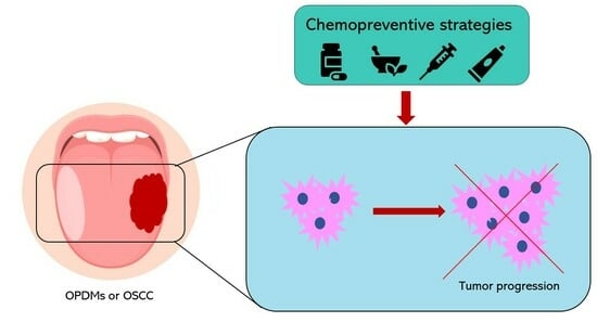
Oral potentially malignant disorders (OPMD) and oral squamous cell carcinoma (OSCC) represent a significant global health burden due to their potential for malignant transformation and the challenges associated with their diagnosis and treatment. Chemoprevention, an innovative approach aimed at halting or reversing the neoplastic process before full malignancy, has emerged as a promising avenue for mitigating the impact of OPMD and OSCC. The pivotal role of chemopreventive strategies is underscored by the need for effective interventions that go beyond traditional therapies. In this regard, chemopreventive agents offer a unique opportunity to intercept disease progression by targeting the molecular pathways implicated in carcinogenesis. Natural compounds, such as curcumin, green tea polyphenols, and resveratrol, exhibit anti-inflammatory, antioxidant, and anti-cancer properties that could make them potential candidates for curtailing the transformation of OPMD to OSCC. Moreover, targeted therapies directed at specific molecular alterations hold promise in disrupting the signaling cascades driving OSCC growth. Immunomodulatory agents, like immune checkpoint inhibitors, are gaining attention for their potential to harness the body’s immune response against early malignancies, thus impeding OSCC advancement. Additionally, nutritional interventions and topical formulations of chemopreventive agents offer localized strategies for preventing carcinogenesis in the oral cavity. The challenge lies in optimizing these strategies for efficacy, safety, and patient compliance. This review presents an up to date on the dynamic interplay between molecular insights, clinical interventions, and the broader goal of reducing the burden of oral malignancies. As research progresses, the synergy between early diagnosis, non-invasive biomarker identification, and chemopreventive therapy is poised to reshape the landscape of OPMD and OSCC management, offering a glimpse of a future where these diseases are no longer insurmountable challenges but rather preventable and manageable conditions.
Full article
(This article belongs to the Special Issue Recent Advances in Drug Delivery and Oral Health: The Impact of Technology and Digital Advances 2.0)
►▼
Show Figures
Graphical abstract
Open AccessArticle
Development of an Anisotropic Hyperelastic Material Model for Porcine Colorectal Tissues
Bioengineering 2024, 11(1), 64; https://doi.org/10.3390/bioengineering11010064 - 08 Jan 2024
Abstract
Many colonic surgeries include colorectal anastomoses whose leaks may be life-threatening, affecting thousands of patients annually. Various studies propose that mechanical interaction between the staples and neighboring tissues may play an important role in anastomotic leakage. Therefore, understanding the mechanical behavior of colorectal
[...] Read more.
Many colonic surgeries include colorectal anastomoses whose leaks may be life-threatening, affecting thousands of patients annually. Various studies propose that mechanical interaction between the staples and neighboring tissues may play an important role in anastomotic leakage. Therefore, understanding the mechanical behavior of colorectal tissue is essential to characterizing the reasons for this type of failure. So far, experimental data characterizing the mechanical properties of colorectal tissue have been few and inconsistent, which has significantly limited understanding their behavior. This research proposes an approach to developing an anisotropic hyperelastic material model for colorectal tissues based on uniaxial testing of freshly harvested porcine specimens, which were collected from several age- and weight-matched pigs. The specimens were extracted from the same colon tract of each pig along their circumferential and longitudinal orientations. We propose a constitutive model combining Yeoh isotropic hyperelastic material with fibers oriented in two directions to account for the hyperelastic and anisotropic nature of colorectal tissues. Experimental data were used to accurately determine the model’s coefficients (circumferential, R2 = 0.9968; longitudinal, R2 = 0.9675). The results show that the proposed model can be incorporated into a finite element model that can simulate procedures such as colorectal anastomoses reliably.
Full article
(This article belongs to the Special Issue Mechanobiology in Biomedical Engineering)
►▼
Show Figures
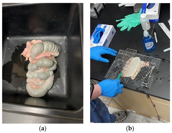
Figure 1
Open AccessCommunication
Neurovascular Relationships in AGEs-Based Models of Proliferative Diabetic Retinopathy
by
, , , , and
Bioengineering 2024, 11(1), 63; https://doi.org/10.3390/bioengineering11010063 - 08 Jan 2024
Abstract
Diabetic retinopathy affects more than 100 million people worldwide and is projected to increase by 50% within 20 years. Increased blood glucose leads to the formation of advanced glycation end products (AGEs), which cause cellular and molecular dysfunction across neurovascular systems. These molecules
[...] Read more.
Diabetic retinopathy affects more than 100 million people worldwide and is projected to increase by 50% within 20 years. Increased blood glucose leads to the formation of advanced glycation end products (AGEs), which cause cellular and molecular dysfunction across neurovascular systems. These molecules initiate the slow breakdown of the retinal vasculature and the inner blood retinal barrier (iBRB), resulting in ischemia and abnormal angiogenesis. This project examined the impact of AGEs in altering the morphology of healthy cells that comprise the iBRB, as well as the effects of AGEs on thrombi formation, in vitro. Our results illustrate that AGEs significantly alter cellular areas and increase the formation of blood clots via elevated levels of tissue factor. Likewise, AGEs upregulate the expression of cell receptors (RAGE) on both endothelial and glial cells, a hallmark biomarker of inflammation in diabetic cells. Examining the effects of AGEs stimulation on cellular functions that work to diminish iBRB integrity will greatly help to advance therapies that target vision loss in adults.
Full article
(This article belongs to the Special Issue Bioengineering and the Eye, Volume II)
►▼
Show Figures
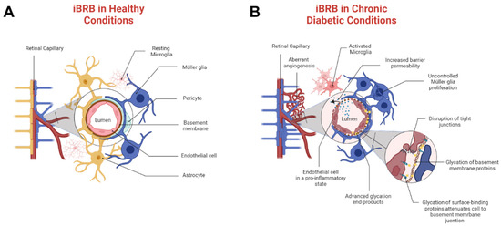
Figure 1

Journal Menu
► ▼ Journal Menu-
- Bioengineering Home
- Aims & Scope
- Editorial Board
- Reviewer Board
- Topical Advisory Panel
- Instructions for Authors
- Special Issues
- Topics
- Sections & Collections
- Article Processing Charge
- Indexing & Archiving
- Editor’s Choice Articles
- Most Cited & Viewed
- Journal Statistics
- Journal History
- Journal Awards
- Society Collaborations
- Conferences
- Editorial Office
Journal Browser
► ▼ Journal BrowserHighly Accessed Articles
Latest Books
E-Mail Alert
News
Topics
Topic in
Energies, Processes, Bioengineering, ChemEngineering, Clean Technol.
Chemical and Biochemical Processes for Energy Sources
Topic Editors: Venko N. Beschkov, Konstantin PetrovDeadline: 31 January 2024
Topic in
Applied Sciences, Bioengineering, Electronics, Healthcare, JCM, Chips
Applied System on Biomedical Engineering, Healthcare and Sustainability 2023
Topic Editors: Teen-Hang Meen, Chun-Yen Chang, Charles Tijus, Po-Lei Lee, Kuei-Shu HsuDeadline: 31 March 2024
Topic in
Bioengineering, Biomolecules, Cancers, Diseases, Nanomaterials, Pharmaceutics
Dynamic Nano-Biomaterials in Tissue Regeneration and Cancer Therapies
Topic Editors: Ramar Thangam, Heemin Kang, Bibin G. Anand, Ramachandran Vijayan, Venugopal KrishnanDeadline: 15 May 2024
Topic in
Applied Sciences, Bioengineering, Fermentation, Processes, Water
Bioreactors: Control, Optimization and Applications - 2nd Volume
Topic Editors: Francesca Raganati, Alessandra ProcenteseDeadline: 30 June 2024

Conferences
Special Issues
Special Issue in
Bioengineering
Advances of Artificial Intelligence for Sustainable Engineering and Medical Applications: Machine Learning and Optimization
Guest Editor: Erfan Babaee TirkolaeeDeadline: 20 January 2024
Special Issue in
Bioengineering
Cognitive Impairment in Multiple Sclerosis
Guest Editors: Elisabetta Signoriello, Cinzia CoppolaDeadline: 31 January 2024
Special Issue in
Bioengineering
Bioinspired and Biogenic Materials for Biomedical and Bioengineering Applications
Guest Editor: Xiang MaDeadline: 15 February 2024
Special Issue in
Bioengineering
Bioanalysis Systems: Materials, Methods, Designs and Applications
Guest Editor: Zongjie WangDeadline: 29 February 2024
Topical Collections
Topical Collection in
Bioengineering
Nanoparticles for Therapeutic and Diagnostic Applications
Collection Editor: Wassana Yantasee
Topical Collection in
Bioengineering
Extracellular Matrix in Engineered Microenvironments for 3D Cell Culture, Tissue Reconstruction, and Disease Modelling In Vitro - 2nd Edition
Collection Editor: Anna E. Guller
Topical Collection in
Bioengineering
Feature Papers in Advanced Computational Technologies for Biosignal Processing
Collection Editors: Yunfeng Wu, Shi Qiu, Behnaz Ghoraani
Topical Collection in
Bioengineering
3D Bioprinting in Bioengineering
Collection Editor: Raymund Horch


