


Biosensors 2024, 14(1), 46; https://doi.org/10.3390/bios14010046 - 15 Jan 2024
Abstract
Cell energy metabolism is a complex and multifaceted process by which some of the most important nutrients, particularly glucose and other sugars, are transformed into energy. This complexity is a result of dynamic interactions between multiple components, including ions, metabolic intermediates, and products
[...] Read more.
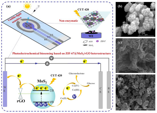
Cell energy metabolism is a complex and multifaceted process by which some of the most important nutrients, particularly glucose and other sugars, are transformed into energy. This complexity is a result of dynamic interactions between multiple components, including ions, metabolic intermediates, and products that arise from biochemical reactions, such as glycolysis and mitochondrial oxidative phosphorylation (OXPHOS), the two main metabolic pathways that provide adenosine triphosphate (ATP), the main source of chemical energy driving various physiological activities. Impaired cell energy metabolism and perturbations or dysfunctions in associated metabolites are frequently implicated in numerous diseases, such as diabetes, cancer, and neurodegenerative and cardiovascular disorders. As a result, altered metabolites hold value as potential disease biomarkers. Electrochemical biosensors are attractive devices for the early diagnosis of many diseases and disorders based on biomarkers due to their advantages of efficiency, simplicity, low cost, high sensitivity, and high selectivity in the detection of anomalies in cellular energy metabolism, including key metabolites involved in glycolysis and mitochondrial processes, such as glucose, lactate, nicotinamide adenine dinucleotide (NADH), reactive oxygen species (ROS), glutamate, and ATP, both in vivo and in vitro. This paper offers a detailed examination of electrochemical biosensors for the detection of glycolytic and mitochondrial metabolites, along with their many applications in cell chips and wearable sensors.
Full article
(This article belongs to the Special Issue Cell-Based Biosensors for Rapid Detection and Monitoring)


