


Dermatopathology 2024, 11(1), 19-25; https://doi.org/10.3390/dermatopathology11010004 - 31 Dec 2023
Abstract
►
Show Figures
Folliculosebaceous cystic hamartoma (FSCH) is a rare and benign form of cutaneous hamartomas. These skin lesions often lead to clinical and histopathological misdiagnosis due to their similarities to cutaneous lesions with overproduction of clustered sebaceous glands. Clinically, the lesions often present as solitary,
[...] Read more.
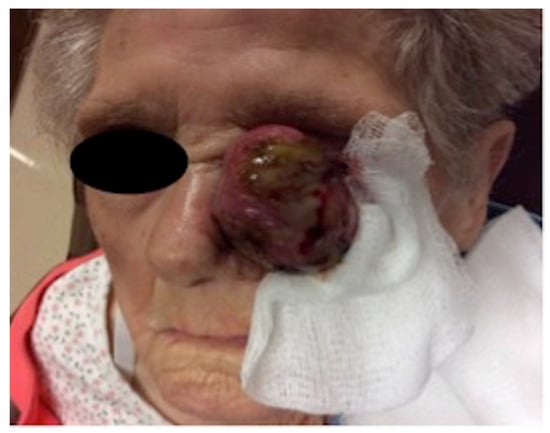
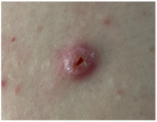
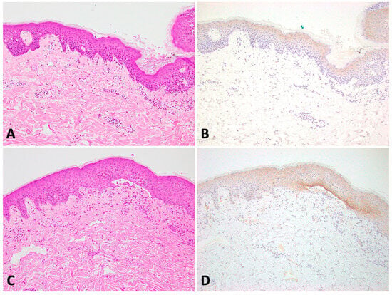
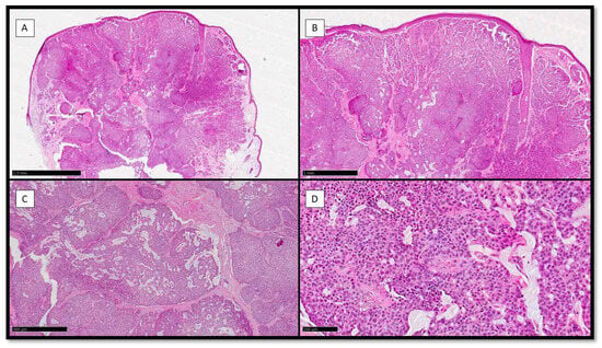
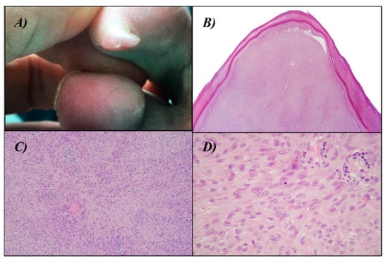
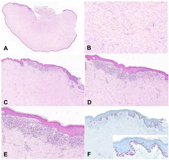
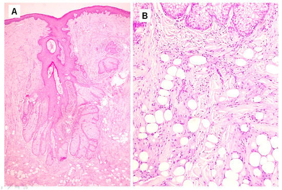
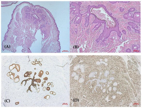
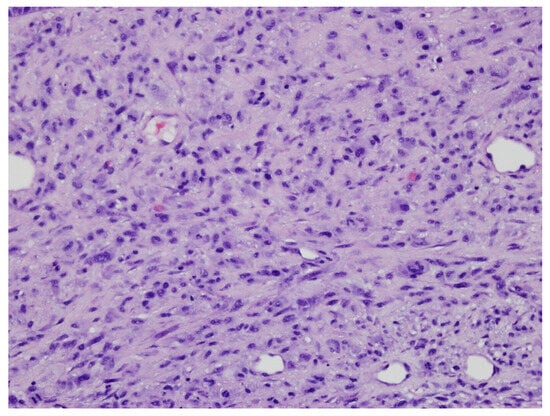
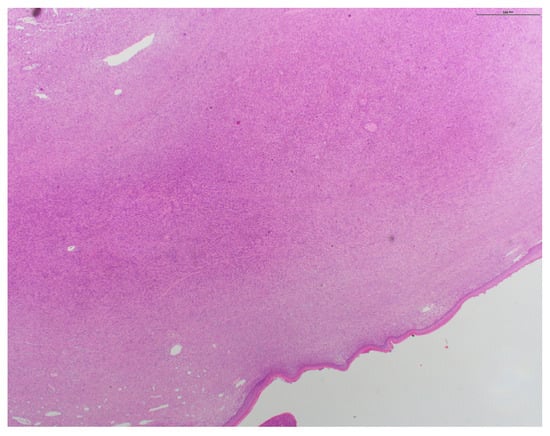
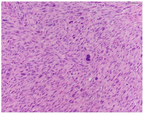
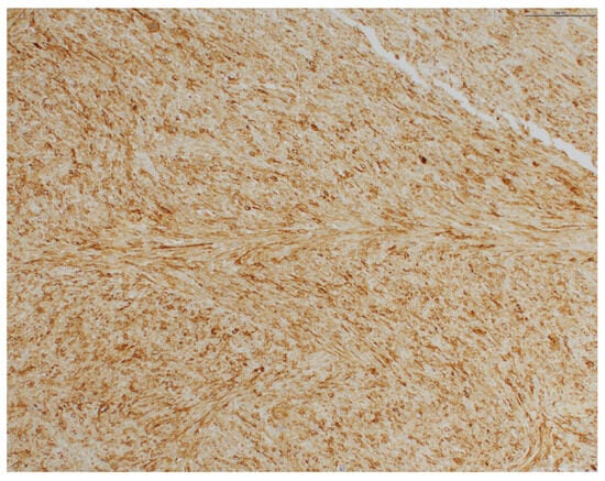
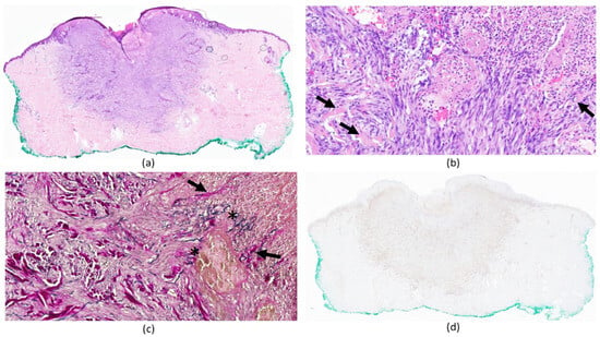
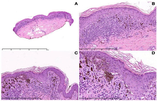
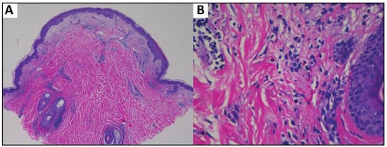
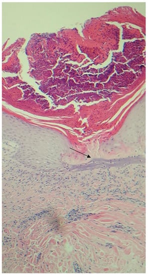
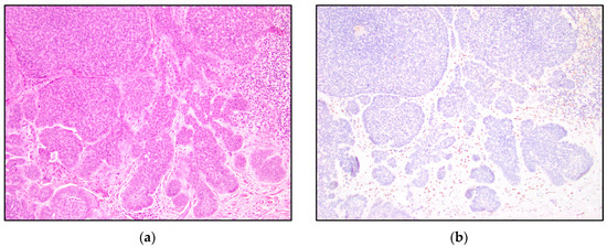
Folliculosebaceous cystic hamartoma (FSCH) is a rare and benign form of cutaneous hamartomas. These skin lesions often lead to clinical and histopathological misdiagnosis due to their similarities to cutaneous lesions with overproduction of clustered sebaceous glands. Clinically, the lesions often present as solitary, skin-colored, pedunculated warts to cauliflower-like, exophytic papules and nodules, usually with a diameter ranging 0.5–1.5 cm that rarely exceed 2 cm in size. Only a small number of giant variants are reported in the literature with a diameter in the range of 5–23 cm. The vast majority of the lesions appear in the central face and show a striking predilection for the nose, ears, and scalp, but also emerge on the nipples, extremities, and genitals. Histologically, the epithelial components of folliculosebaceous cystic hamartoma comprise dilated infundibular cystic proliferation with surrounding mesenchymal components, which commonly include fibroplasia and vascular and adipose tissue proliferation. These histological characteristics were coined by Kimura and colleagues (1991). To the best of our knowledge, our case represents the biggest variant of giant folliculosebaceous cystic hamartoma.
Full article
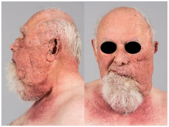
Figure 1


