


by
, , , , , , , , and

Diagnostics 2024, 14(2), 190; https://doi.org/10.3390/diagnostics14020190 - 15 Jan 2024
Abstract
Volitional assessment of quadriceps muscle endurance is clinically relevant in patients with chronic obstructive pulmonary disease (COPD). However, studies that determine the construct validity of volitional tests by comparing them to non-volitional measures are lacking. Therefore, the aim of the current study is
[...] Read more.
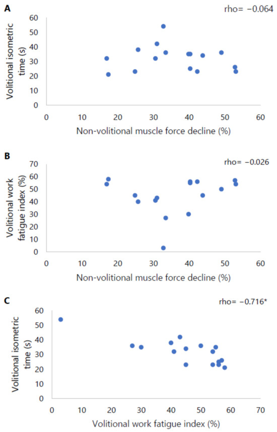
Volitional assessment of quadriceps muscle endurance is clinically relevant in patients with chronic obstructive pulmonary disease (COPD). However, studies that determine the construct validity of volitional tests by comparing them to non-volitional measures are lacking. Therefore, the aim of the current study is to evaluate the correlation between volitional and non-volitional quadriceps muscle endurance in patients with COPD. Quadriceps muscle endurance was evaluated in twenty-six patients with COPD. A volitional isometric and a volitional isokinetic protocol were performed on a computerised dynamometer to determine the isometric time and isokinetic work fatigue index, respectively. Non-volitional assessment of quadriceps muscle endurance was evaluated using repetitive electrical stimulations to establish the isometric muscle force decline. Sixteen patients (61 ± 8 years, 63% male, FEV1 47 (32–53)%) performed all three quadriceps endurance tests conforming to pre-defined test criteria. Both volitional isometric time and isokinetic work fatigue index did not significantly correlate with non-volitional muscle force decline (both p > 0.05). There was a strong correlation between volitional isometric time and isokinetic work fatigue index (rho = −0.716, p = 0.002). To conclude, this study suggests that volitional measures evaluate partly different aspects of quadriceps muscle endurance compared to non-volitional measures. Accordingly, these outcome measures cannot be used interchangeably.
Full article
(This article belongs to the Special Issue Technologies in the Diagnosis of Lung Diseases)
►
Show Figures
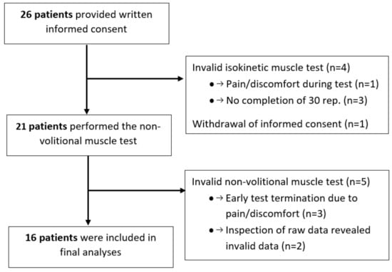
Figure 1


.JPG)


