


J. Dev. Biol. 2024, 12(1), 4; https://doi.org/10.3390/jdb12010004 - 15 Jan 2024
Abstract
The epidermal differentiation complex (EDC) is a cluster of genes that encode protein components of the outermost layers of the epidermis in mammals, reptiles and birds. The development of the stratified epidermis from a single-layered ectoderm involves an embryo-specific superficial cell layer, the
[...] Read more.
The epidermal differentiation complex (EDC) is a cluster of genes that encode protein components of the outermost layers of the epidermis in mammals, reptiles and birds. The development of the stratified epidermis from a single-layered ectoderm involves an embryo-specific superficial cell layer, the periderm. An additional layer, the subperiderm, develops in crocodilians and over scutate scales of birds. Here, we review the expression of EDC genes during embryonic development. Several EDC genes are expressed predominantly or exclusively in embryo-specific cell layers, whereas others are confined to the epidermal layers that are maintained in postnatal skin. The S100 fused-type proteins scaffoldin and trichohyalin are expressed in the avian and mammalian periderm, respectively. Scaffoldin forms the so-called periderm granules, which are histological markers of the periderm in birds. Epidermal differentiation cysteine-rich protein (EDCRP) and epidermal differentiation protein containing DPCC motifs (EDDM) are expressed in the avian subperiderm where they are supposed to undergo cross-linking via disulfide bonds. Furthermore, a histidine-rich epidermal differentiation protein and feather-type corneous beta-proteins, also known as beta-keratins, are expressed in the subperiderm. The accumulating evidence for roles of EDC genes in the development of the epidermis has implications on the evolutionary diversification of the skin in amniotes.
Full article
(This article belongs to the Special Issue The 10th Anniversary of JDB: Feature Papers)
►
Show Figures
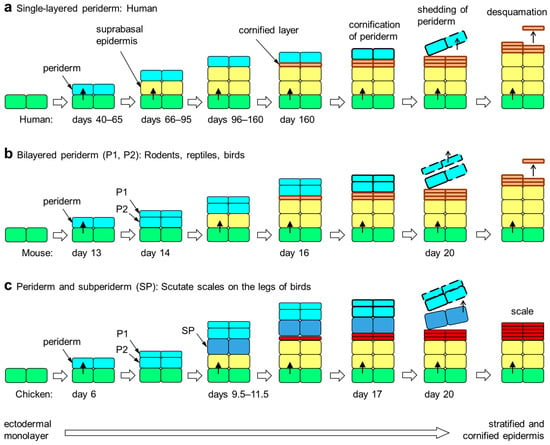
Figure 1


