
Lymphatics 2024, 2(1), 10-24; https://doi.org/10.3390/lymphatics2010002 - 05 Jan 2024
Abstract
Anti-CD20 monoclonal antibodies (mAbs) have revolutionized the treatment of lymphomas by improving the survival of patients, particularly in conjunction with chemotherapy. Until recently, the gold standard was based on the utilization of Rituximab (RTX) combined with chemotherapy. With our better understanding of monoclonal
[...] Read more.
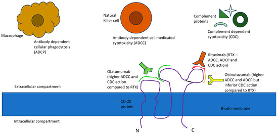
Anti-CD20 monoclonal antibodies (mAbs) have revolutionized the treatment of lymphomas by improving the survival of patients, particularly in conjunction with chemotherapy. Until recently, the gold standard was based on the utilization of Rituximab (RTX) combined with chemotherapy. With our better understanding of monoclonal antibody (mAb) engineering, anti-CD20 mAb therapy has evolved to enhance clinical outcomes by improving pharmacokinetics, safety, activity and immunogenicity. Efforts to improve the on-targeting CD20 expressed on lymphomas through novel bioengineering techniques have led to the development of newer anti-CD20 mAbs that have accentuated complement-dependent cytotoxicity (CDC), antibody-dependent cell medicated cytotoxicity (ADCC), and/or a direct killing effect. There are several anti-CD20 monoclonal antibodies that have been evaluated for the treatment of lymphomas, some of which are now approved in addition to RTX.
Full article
(This article belongs to the Collection Lymphomas)
►
Show Figures
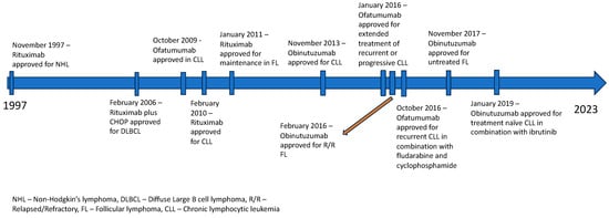
Figure 1


